Copyright
©The Author(s) 2023.
World J Gastrointest Surg. Mar 27, 2023; 15(3): 408-419
Published online Mar 27, 2023. doi: 10.4240/wjgs.v15.i3.408
Published online Mar 27, 2023. doi: 10.4240/wjgs.v15.i3.408
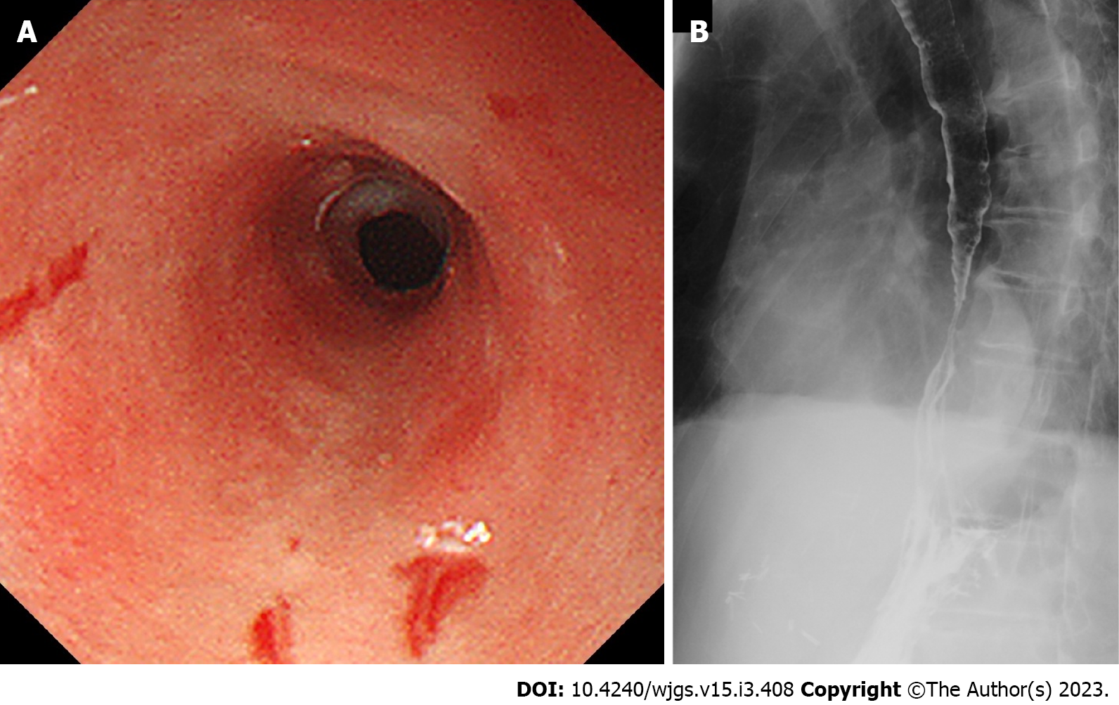
Figure 6 A case of esophageal stenosis.
A 75-year-old male patient developed non-black esophagus acute esophageal mucosal lesions during chemotherapy for colorectal cancer. The patient could not eat on the 27th day of onset. Upper endoscopy and esophagography showed esophageal stenosis. Symptoms improved after five endoscopic balloon dilatations. A: Upper endoscopy; B: Esophagography.
- Citation: Ichita C, Sasaki A, Shimizu S. Clinical features of acute esophageal mucosal lesions and reflux esophagitis Los Angeles classification grade D: A retrospective study. World J Gastrointest Surg 2023; 15(3): 408-419
- URL: https://www.wjgnet.com/1948-9366/full/v15/i3/408.htm
- DOI: https://dx.doi.org/10.4240/wjgs.v15.i3.408


