Copyright
©The Author(s) 2023.
World J Gastrointest Surg. Mar 27, 2023; 15(3): 398-407
Published online Mar 27, 2023. doi: 10.4240/wjgs.v15.i3.398
Published online Mar 27, 2023. doi: 10.4240/wjgs.v15.i3.398
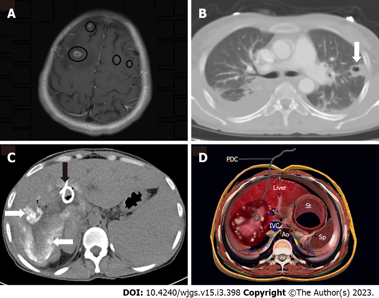
Figure 4 Male, 45 years old.
A and B: Axial magnetic resonance imaging demonstrates enhancing brain lesions associated with alveolar echinococcosis (circles). Alveolar Echinococcosis is also prevalent in the parenchyma of the left lung (arrow); C: Percutaneous drainage was performed; typical calcifications (white arrow) are visible, as is the drainage catheter (black arrow); D: 3D axial plane cross sectional illustration image shows the percutaneous drainage catheter placement in the lesion cavity. PDC: Percutaneous drainage catheter; IVC: Inferior vena cava; Ao: Abdominal aorta; IVC: Inferior vena cava; St: Stomach; Sp: Spleen.
- Citation: Eren S, Aydın S, Kantarci M, Kızılgöz V, Levent A, Şenbil DC, Akhan O. Percutaneous management in hepatic alveolar echinococcosis: A sum of single center experiences and a brief overview of the literature. World J Gastrointest Surg 2023; 15(3): 398-407
- URL: https://www.wjgnet.com/1948-9366/full/v15/i3/398.htm
- DOI: https://dx.doi.org/10.4240/wjgs.v15.i3.398


