Copyright
©The Author(s) 2023.
World J Gastrointest Surg. Mar 27, 2023; 15(3): 398-407
Published online Mar 27, 2023. doi: 10.4240/wjgs.v15.i3.398
Published online Mar 27, 2023. doi: 10.4240/wjgs.v15.i3.398
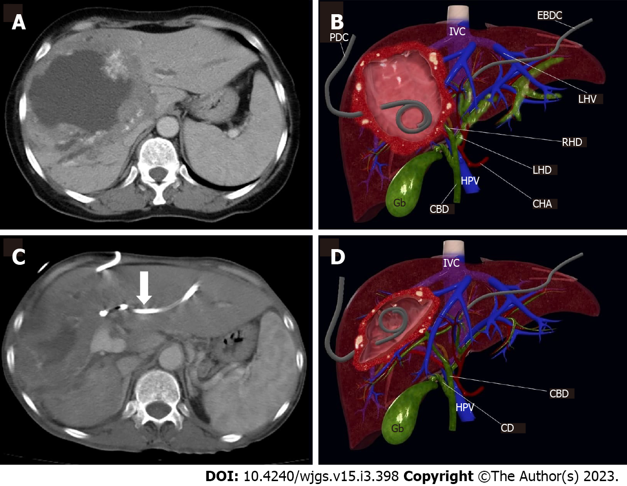
Figure 3 Female, 32-year-old.
A: Alveolar Echinococcosis at right liver lobe, with typical peripheral calcifications and large central necrosis; B: 3D image in the coronal plane illustrates the location of the percutaneous drainage catheter and the external biliary drainage catheter in the patient; C: Drainage catheter can be seen in within the lesion (arrow); D: The shrunken lesion cavity and the regression of the dilatation in the left intrahepatic bile ducts are illustrated by 3D coronal plane image. PDC: Percutaneous drainage catheter; IVC: Inferior vena cava; EBDC: External biliary drainage catheter; LHV: Left hepatic vein; RHD: Right hepatic duct; LHD: Left hepatic duct; CHA: Common hepatic artery; HPV: Hepatic portal vein; CBD: Common biliary duct; Gb: Gallbladder; CD: Cystic duct.
- Citation: Eren S, Aydın S, Kantarci M, Kızılgöz V, Levent A, Şenbil DC, Akhan O. Percutaneous management in hepatic alveolar echinococcosis: A sum of single center experiences and a brief overview of the literature. World J Gastrointest Surg 2023; 15(3): 398-407
- URL: https://www.wjgnet.com/1948-9366/full/v15/i3/398.htm
- DOI: https://dx.doi.org/10.4240/wjgs.v15.i3.398


