Copyright
©The Author(s) 2023.
World J Gastrointest Surg. Feb 27, 2023; 15(2): 294-302
Published online Feb 27, 2023. doi: 10.4240/wjgs.v15.i2.294
Published online Feb 27, 2023. doi: 10.4240/wjgs.v15.i2.294
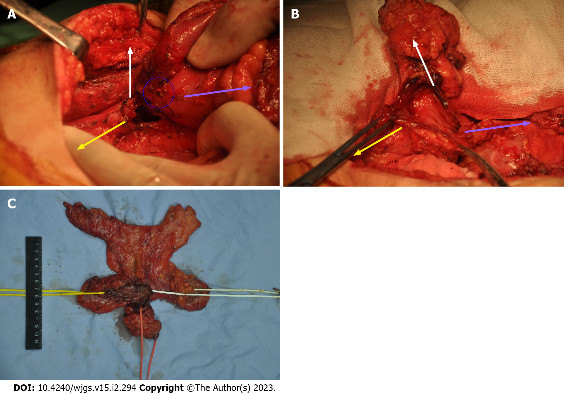
Figure 4 The structure of the t-branch tube.
A: The yellow arrow indicates the proximal colon, the white arrow indicates the colostomy colon, the purple arrow indicates the distal colon, and the blue circle indicates the small intestinal wall. Intraoperative exploration confirmed that the t-branch tube was composed of the distal colon, proximal colon, colostomy colon, and small intestinal wall; B: After separating the small intestinal wall, the structure of the t-branch tube could be more clearly identified; C: Surgical removal of the t-branch tube structure of the colon. The yellow marker shows the proximal colon, the green marker indicates the distal colon, the orange marker shows the original stoma, and the defect is the original small intestinal wall.
- Citation: Zhang Y, Lin H, Liu JM, Wang X, Cui YF, Lu ZY. Mesh erosion into the colon following repair of parastomal hernia: A case report. World J Gastrointest Surg 2023; 15(2): 294-302
- URL: https://www.wjgnet.com/1948-9366/full/v15/i2/294.htm
- DOI: https://dx.doi.org/10.4240/wjgs.v15.i2.294


