Copyright
©The Author(s) 2023.
World J Gastrointest Surg. Feb 27, 2023; 15(2): 193-200
Published online Feb 27, 2023. doi: 10.4240/wjgs.v15.i2.193
Published online Feb 27, 2023. doi: 10.4240/wjgs.v15.i2.193
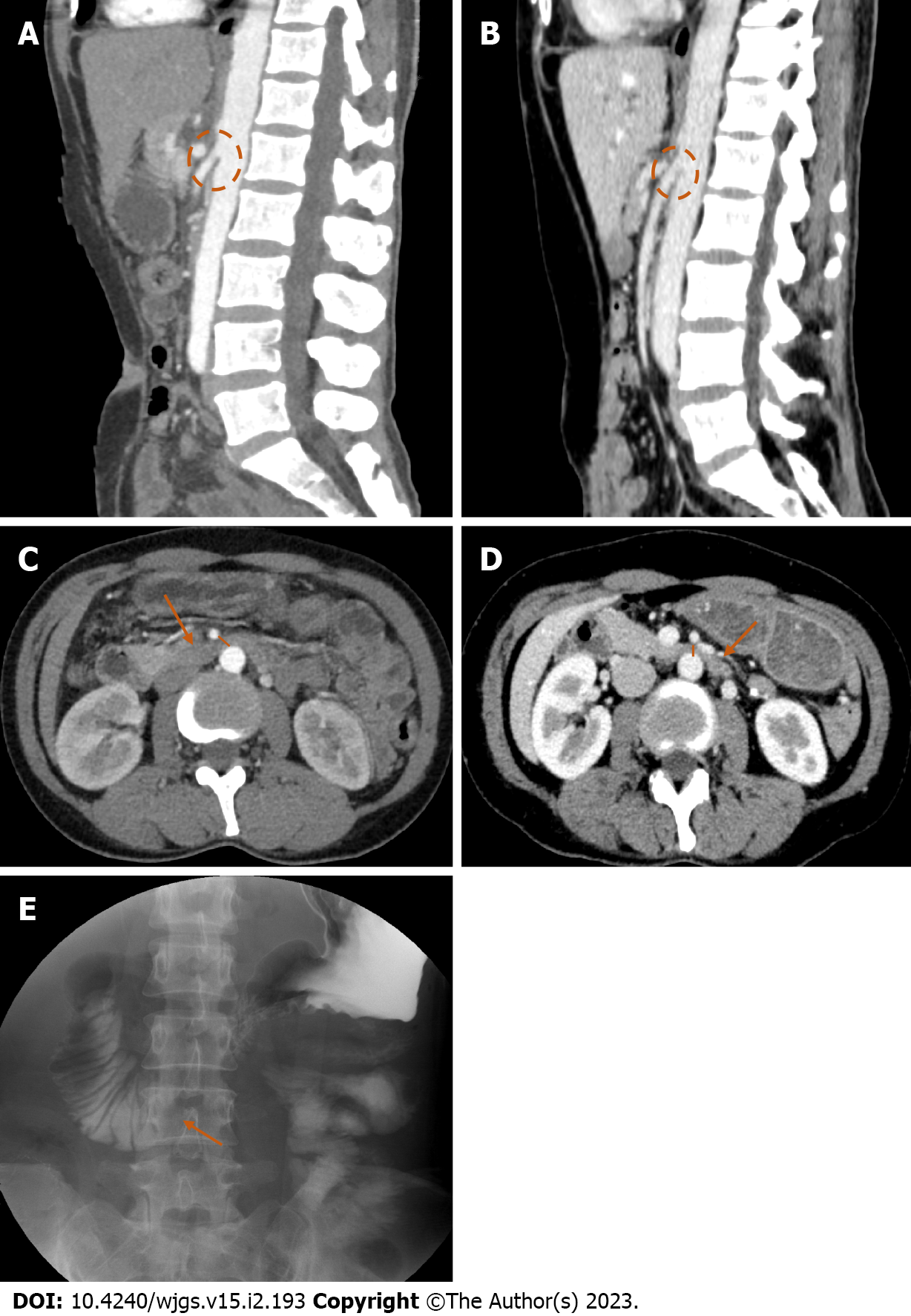
Figure 1 Imaging of a patient with postoperative superior mesenteric artery syndrome.
A: Abdominal enhanced computed tomography (CT) in the preoperative period showing the angle between the superior mesenteric artery (SMA) and abdominal aorta (AA) was 19°; B: Abdominal enhanced CT in the postoperative period showing the altered anatomical position of the SMA and the aortomesenteric angle was 10°; C: Abdominal enhanced CT in the preoperative period showing the distance between the SMA and AA in the third portion (large arrow) of the duodenum was 7 mm (short thin line); D: Abdominal enhanced CT in the postoperative period showing the reduced aortmesenteric distance in the third portion (large arrow) was 4 mm (short thin line); E: Representative image from upper gastrointestinal series showing abrupt cutoff of oral contrast in the third portion of the duodenum (arrow) and slight dilation of the proximal duodenum, suggestive of superior mesenteric artery syndrome.
- Citation: Xie J, Bai J, Zheng T, Shu J, Liu ML. Causes of epigastric pain and vomiting after laparoscopic-assisted radical right hemicolectomy - superior mesenteric artery syndrome. World J Gastrointest Surg 2023; 15(2): 193-200
- URL: https://www.wjgnet.com/1948-9366/full/v15/i2/193.htm
- DOI: https://dx.doi.org/10.4240/wjgs.v15.i2.193


