Copyright
©The Author(s) 2023.
World J Gastrointest Surg. Dec 27, 2023; 15(12): 2945-2953
Published online Dec 27, 2023. doi: 10.4240/wjgs.v15.i12.2945
Published online Dec 27, 2023. doi: 10.4240/wjgs.v15.i12.2945
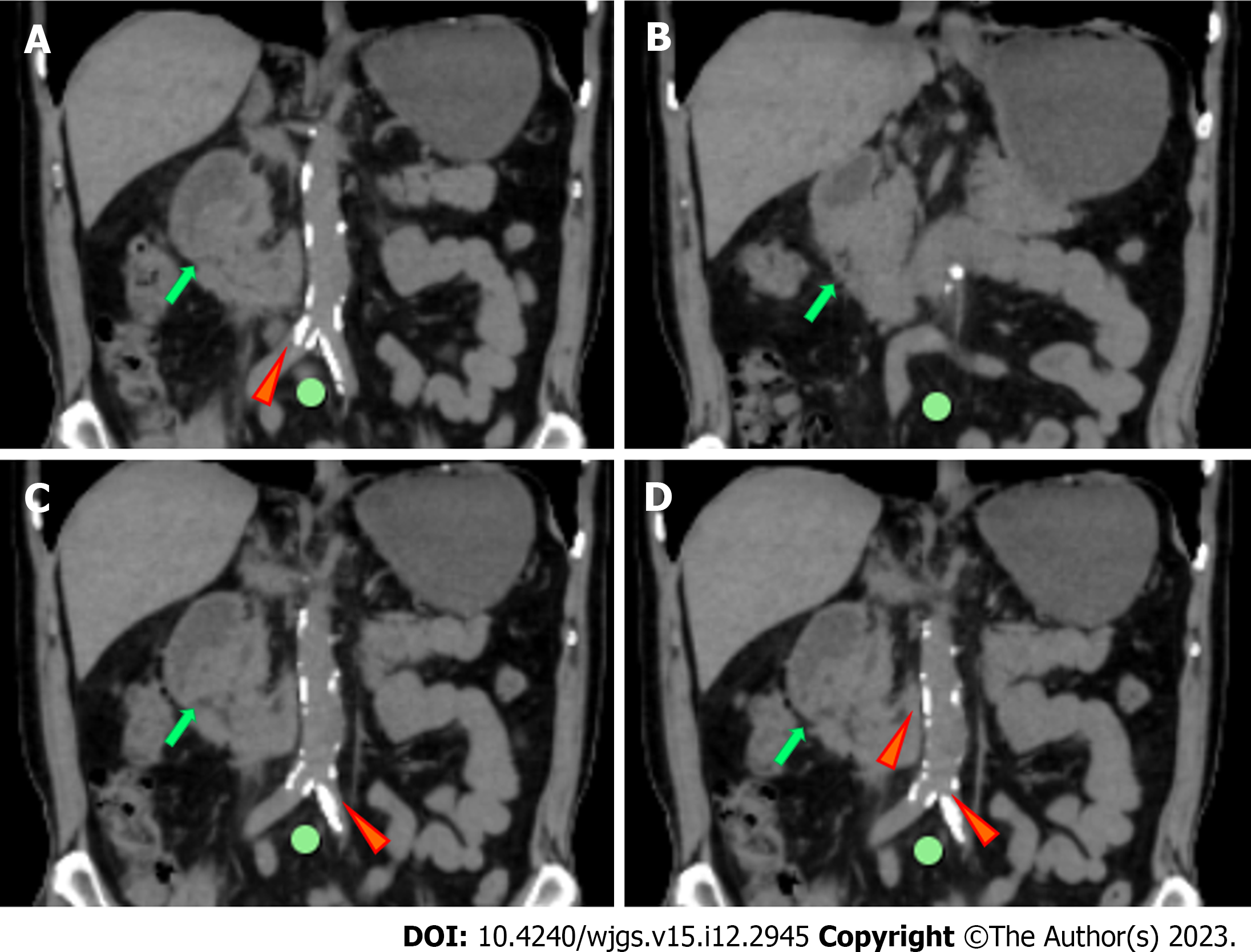
Figure 2 Abdominal plain computed tomography, coronal view.
A-D: An irregular nodule was found in the descending part of the duodenum with no clear demarcation (green arrows); A, C and D: Images showed abdominal aortic wall calcification (red arrows) and partial calcified endometrium inward migration.
- Citation: Zhang Y, Cheng HH, Fan WJ. Duodenojejunostomy treatment of groove pancreatitis-induced stenosis and obstruction of the horizontal duodenum: A case report. World J Gastrointest Surg 2023; 15(12): 2945-2953
- URL: https://www.wjgnet.com/1948-9366/full/v15/i12/2945.htm
- DOI: https://dx.doi.org/10.4240/wjgs.v15.i12.2945


