Copyright
©The Author(s) 2023.
World J Gastrointest Surg. Dec 27, 2023; 15(12): 2945-2953
Published online Dec 27, 2023. doi: 10.4240/wjgs.v15.i12.2945
Published online Dec 27, 2023. doi: 10.4240/wjgs.v15.i12.2945
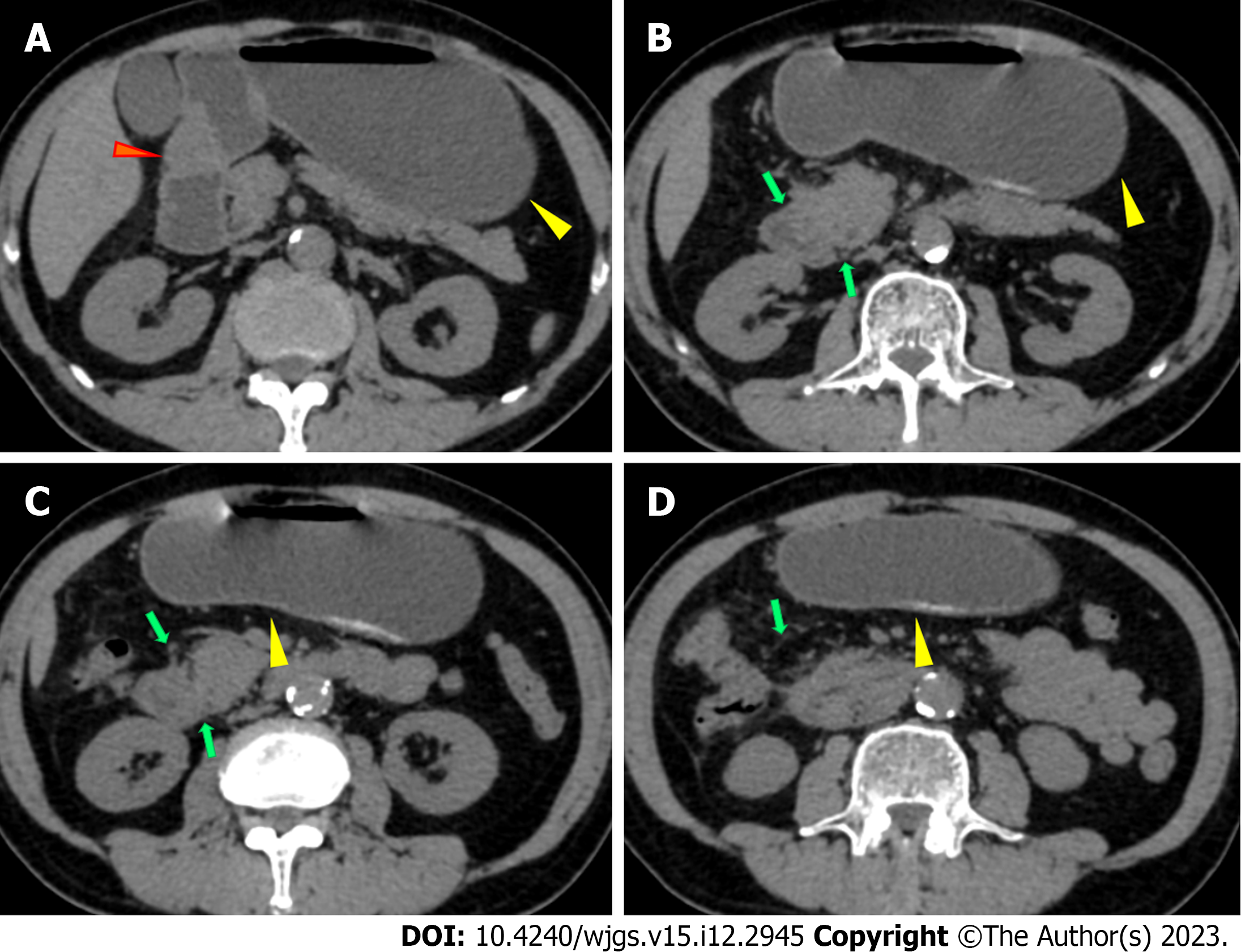
Figure 1 Abdominal plain computed tomography, axial view.
A: Abdominal plain computed tomography (CT) showed gastric retention (yellow arrow) and a nodule (35 mm × 23 mm) in the anterior wall of the pylorus (red arrow); B-D: Abdominal CT showed slight exudation in the descending and horizontal parts of the duodenum with broadening of the groove region, indicating local pancreatitis around the pancreatic head (green arrows) and gastric retention (yellow arrows).
- Citation: Zhang Y, Cheng HH, Fan WJ. Duodenojejunostomy treatment of groove pancreatitis-induced stenosis and obstruction of the horizontal duodenum: A case report. World J Gastrointest Surg 2023; 15(12): 2945-2953
- URL: https://www.wjgnet.com/1948-9366/full/v15/i12/2945.htm
- DOI: https://dx.doi.org/10.4240/wjgs.v15.i12.2945


