Copyright
©The Author(s) 2023.
World J Gastrointest Surg. Dec 27, 2023; 15(12): 2926-2931
Published online Dec 27, 2023. doi: 10.4240/wjgs.v15.i12.2926
Published online Dec 27, 2023. doi: 10.4240/wjgs.v15.i12.2926
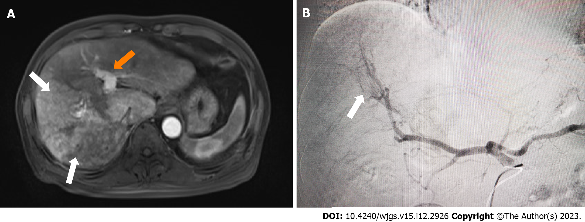
Figure 1 A 58-year-old man with hepatocellular carcinoma and marked arterioportal shunt due to portal vein tumor thrombus.
A: Axial magnetic resonance imaging T1 post contrast weighted image showing a large hypervascular mass in the right hepatic lobe (white arrows). Note enhancement of the portal vein (orange arrow) in the arterial phase denoting an underlying arterioportal shunt; B: Digital subtraction angiography showing opacified portal vein (white arrows) during the early arterial phase.
- Citation: Wang XD, Ge NJ, Yang YF. Portal vein embolization for closure of marked arterioportal shunt of hepatocellular carcinoma to enable radioembolization: A case report. World J Gastrointest Surg 2023; 15(12): 2926-2931
- URL: https://www.wjgnet.com/1948-9366/full/v15/i12/2926.htm
- DOI: https://dx.doi.org/10.4240/wjgs.v15.i12.2926


