Copyright
©The Author(s) 2023.
World J Gastrointest Surg. Nov 27, 2023; 15(11): 2663-2673
Published online Nov 27, 2023. doi: 10.4240/wjgs.v15.i11.2663
Published online Nov 27, 2023. doi: 10.4240/wjgs.v15.i11.2663
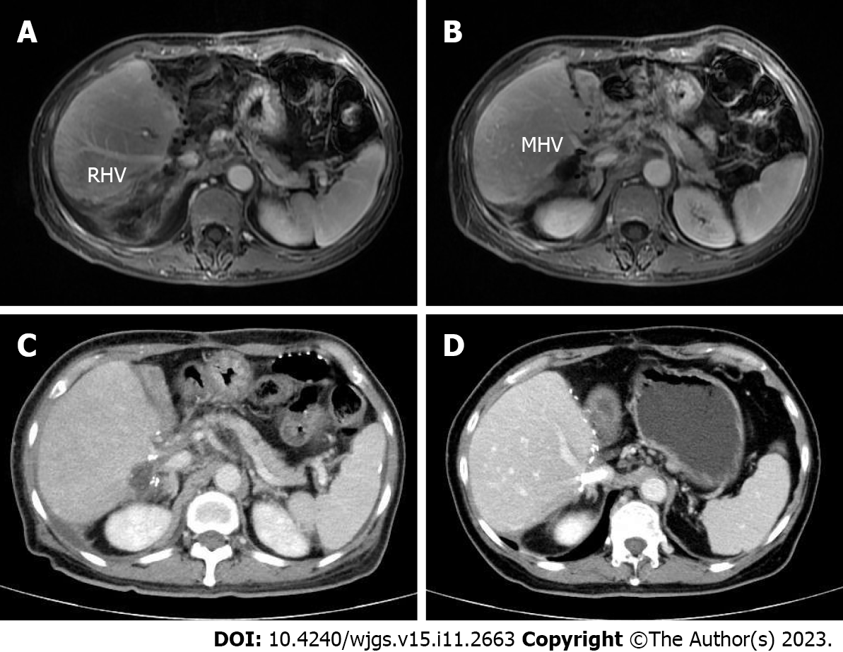
Figure 5 Contrast-enhanced magnetic resonance images and computed tomography images after surgery.
A and B: Portal venous phase image after surgery clearly showing the middle hepatic vein and right portal vein, respectively; C: Portal venous phase image showing the existence of hepatic congestion and hepatomegaly; D: Portal venous phase image showing the hepatic vein stent. MHV: Middle hepatic vein; RHV: Right hepatic vein.
- Citation: Hu CL, Han X, Gao ZZ, Zhou B, Tang JL, Pei XR, Lu JN, Xu Q, Shen XP, Yan S, Ding Y. Systematic sequential therapy for ex vivo liver resection and autotransplantation: A case report and review of literature. World J Gastrointest Surg 2023; 15(11): 2663-2673
- URL: https://www.wjgnet.com/1948-9366/full/v15/i11/2663.htm
- DOI: https://dx.doi.org/10.4240/wjgs.v15.i11.2663


