Copyright
©The Author(s) 2023.
World J Gastrointest Surg. Nov 27, 2023; 15(11): 2513-2524
Published online Nov 27, 2023. doi: 10.4240/wjgs.v15.i11.2513
Published online Nov 27, 2023. doi: 10.4240/wjgs.v15.i11.2513
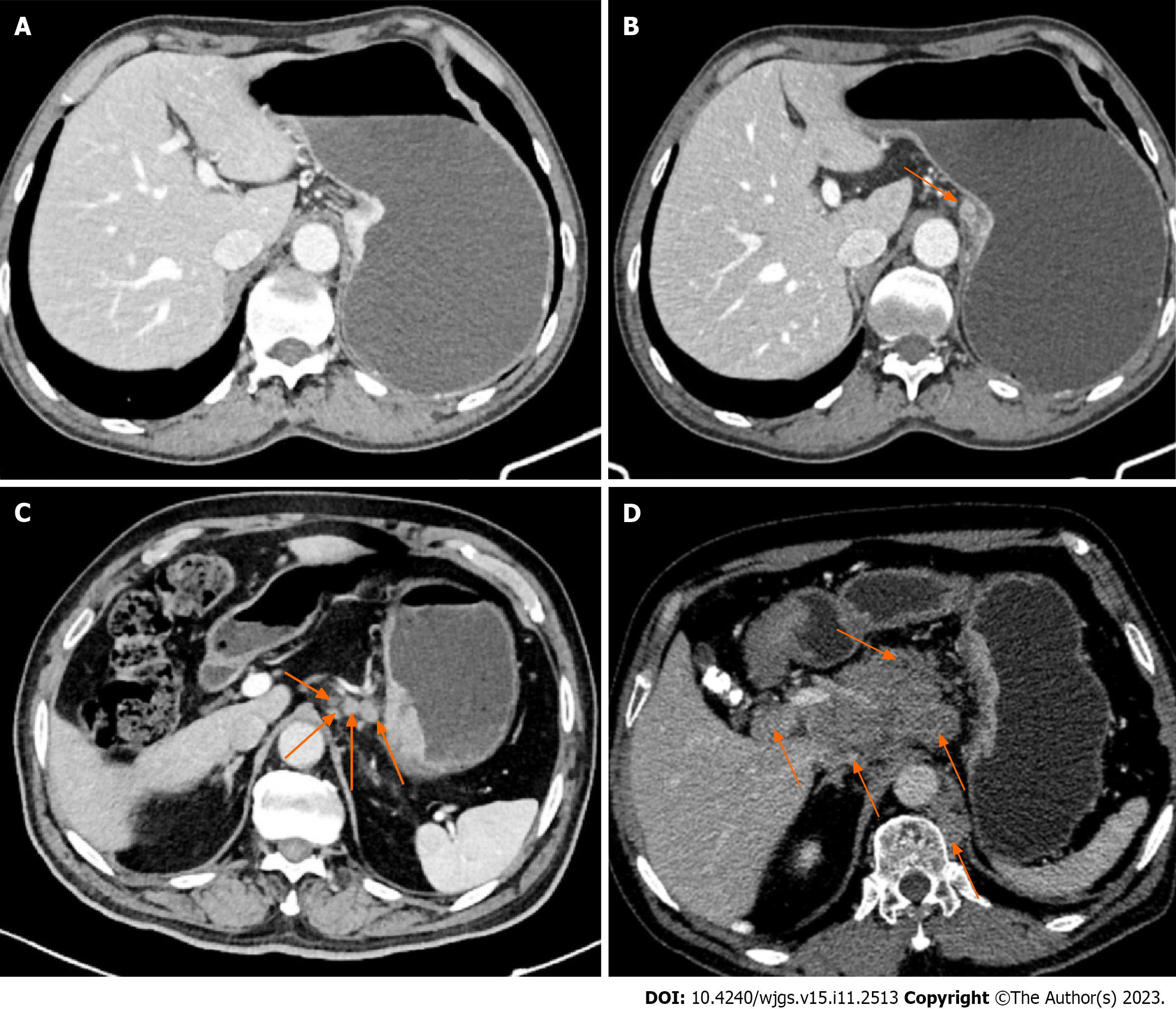
Figure 2 N staging based on three-phase dynamic contrast-enhanced computed tomography scanning.
A: N0: No local lymph node metastasis; B: N1: On the lesser curvature of the stomach, an enlarged lymph node with a diameter of approximately 10 mm is quasi-round, and slightly inhomogeneous enhancement can be observed (orange arrow); C: N2: More than 3 local lymph node metastases were in the left cardia, right cardia, and lesser curvature of the stomach, and the largest was located in the lesser curvature of the stomach, with a short diameter of approximately 12 mm (orange arrows); D: N3: There were more than 7 local lymph node metastases (e.g., porta hepatic, common hepatic artery, left gastric artery, splenic artery, celiac trunk), the lymph nodes were fused into clusters, and the lymph nodes were necrotic and uneven enhancement (orange arrows).
- Citation: Liu H, Zhao KY. Application of CD34 expression combined with three-phase dynamic contrast-enhanced computed tomography scanning in preoperative staging of gastric cancer. World J Gastrointest Surg 2023; 15(11): 2513-2524
- URL: https://www.wjgnet.com/1948-9366/full/v15/i11/2513.htm
- DOI: https://dx.doi.org/10.4240/wjgs.v15.i11.2513


