Copyright
©The Author(s) 2023.
World J Gastrointest Surg. Oct 27, 2023; 15(10): 2108-2114
Published online Oct 27, 2023. doi: 10.4240/wjgs.v15.i10.2108
Published online Oct 27, 2023. doi: 10.4240/wjgs.v15.i10.2108
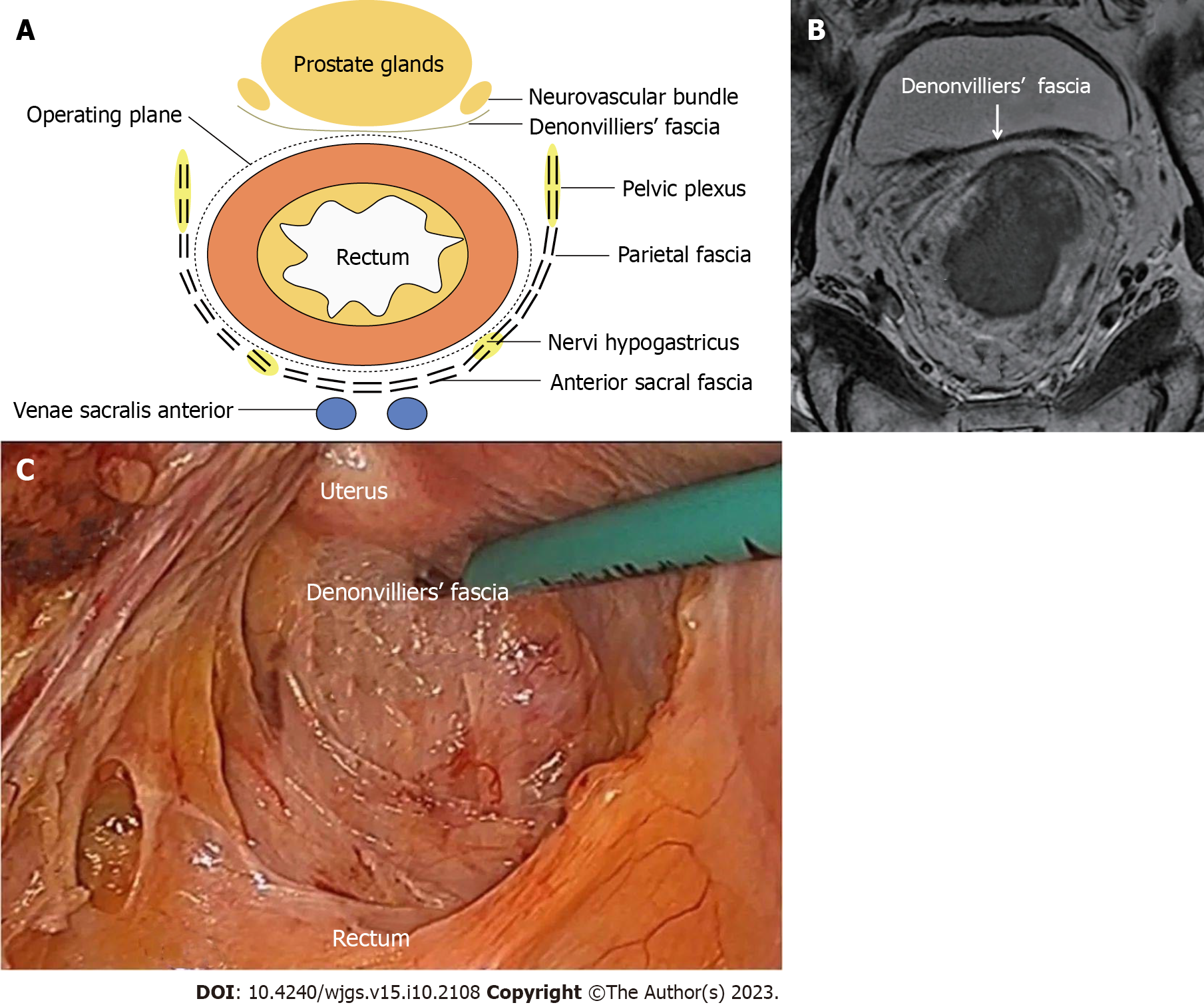
Figure 1 The anatomical location of the Denonvilliers' fascia is shown in hand-drawn diagrams, images and surgical photographs.
A: Diagram of the anatomical pattern of Denonvilliers' fascia (DVF); B: Magnetic resonance imaging-weighted image with DVF shown by arrow; C: Anatomical location and adjacency of DVF during laparoscopy.
- Citation: Chen Z, Zhang XJ, Chang HD, Chen XQ, Liu SS, Wang W, Chen ZH, Ma YB, Wang L. From basic to clinical: Anatomy of Denonvilliers’ fascia and its application in laparoscopic radical resection of rectal cancer. World J Gastrointest Surg 2023; 15(10): 2108-2114
- URL: https://www.wjgnet.com/1948-9366/full/v15/i10/2108.htm
- DOI: https://dx.doi.org/10.4240/wjgs.v15.i10.2108


