Copyright
©The Author(s) 2022.
World J Gastrointest Surg. Aug 27, 2022; 14(8): 731-742
Published online Aug 27, 2022. doi: 10.4240/wjgs.v14.i8.731
Published online Aug 27, 2022. doi: 10.4240/wjgs.v14.i8.731
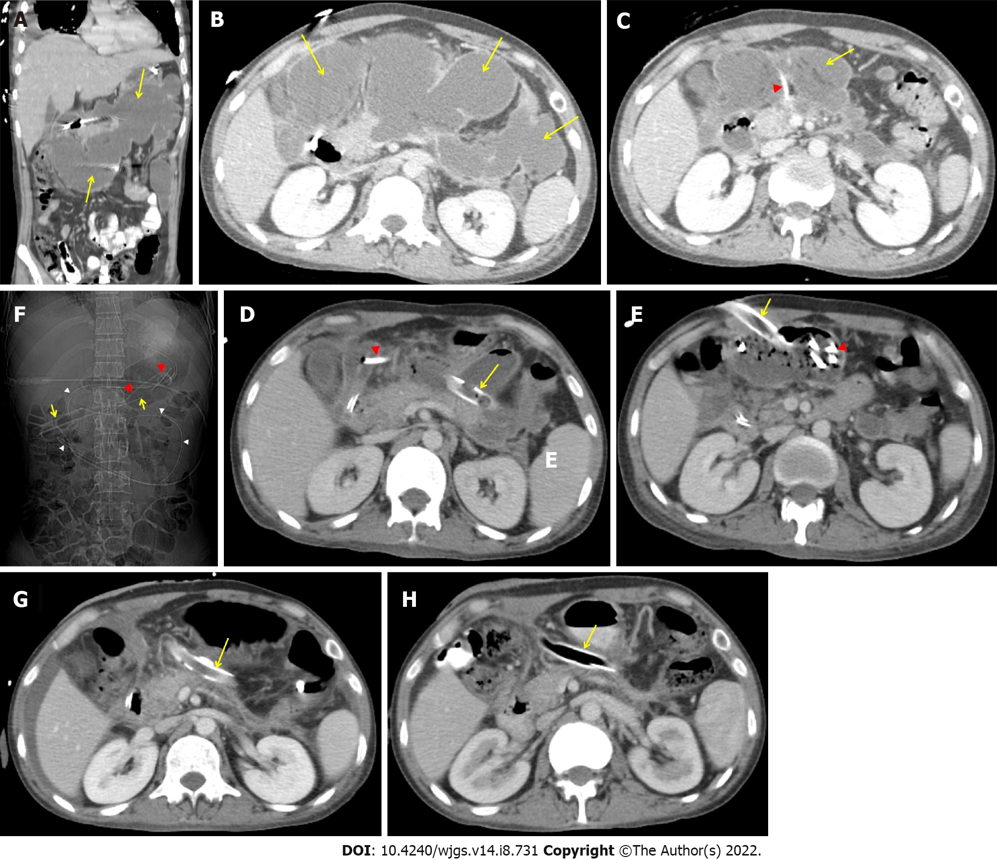
Figure 2 Abdominal contrast enhanced computerized tomography.
A and B: Large, irregular infected pancreatic/peripancreatic collection (PFC) (arrows) in upper abdomen in coronal and transverse sections; C: Partial resolution of PFC (arrow) with a 14 F pigtail (arrow head) in situ; D-F: A 26 F drain (arrows) and a 7 F pigtail irrigation catheter (red arrow head) in walled off pancreatic necrosis (WOPN), and nasojejunal tube (white arrow heads); G and H: A 32 F drain (arrow) in situ with complete resolution of WOPN after (G) 2 wk and (H) 4 wk of percutaneous direct endoscopic necrosectomy.
- Citation: Vyawahare MA, Gulghane S, Titarmare R, Bawankar T, Mudaliar P, Naikwade R, Timane JM. Percutaneous direct endoscopic pancreatic necrosectomy. World J Gastrointest Surg 2022; 14(8): 731-742
- URL: https://www.wjgnet.com/1948-9366/full/v14/i8/731.htm
- DOI: https://dx.doi.org/10.4240/wjgs.v14.i8.731


