Copyright
©The Author(s) 2022.
World J Gastrointest Surg. Jul 27, 2022; 14(7): 656-669
Published online Jul 27, 2022. doi: 10.4240/wjgs.v14.i7.656
Published online Jul 27, 2022. doi: 10.4240/wjgs.v14.i7.656
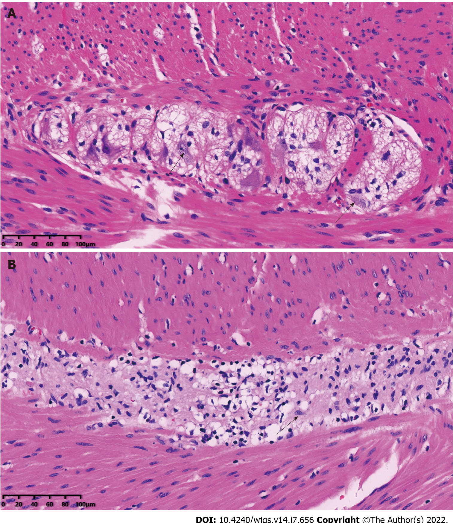
Figure 4 Typical pathological sections of normal intestinal ganglion and resection bowel of allied disorders of patients with Hirschsprung’s disease (hematoxylin-eosin staining).
A: Black arrows indicate normal ganglion cells; B: The black arrow indicates the degenerated ganglion cell. The proliferation of nerve fibers and reduction of ganglion cells were also observed. Magnification, × 200.
- Citation: Jiang S, Song CY, Feng MX, Lu YQ. Adult patients with allied disorders of Hirschsprung’s disease in emergency department: An 11-year retrospective study. World J Gastrointest Surg 2022; 14(7): 656-669
- URL: https://www.wjgnet.com/1948-9366/full/v14/i7/656.htm
- DOI: https://dx.doi.org/10.4240/wjgs.v14.i7.656


