Copyright
©The Author(s) 2022.
World J Gastrointest Surg. Apr 27, 2022; 14(4): 370-373
Published online Apr 27, 2022. doi: 10.4240/wjgs.v14.i4.370
Published online Apr 27, 2022. doi: 10.4240/wjgs.v14.i4.370
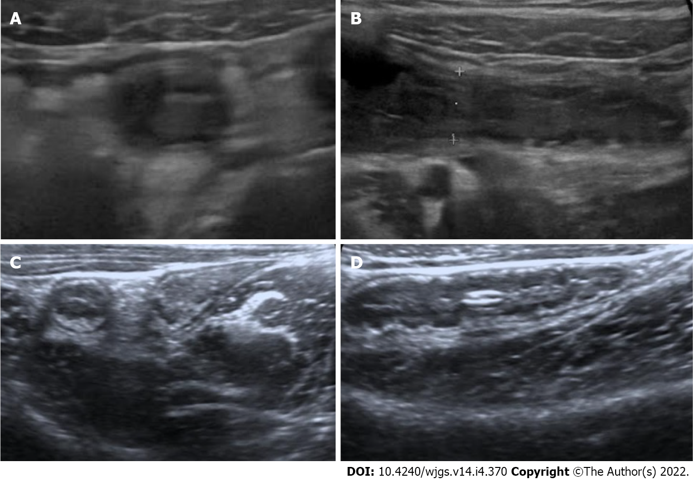
Figure 2 Acute appendicitis in a 12-yr-old boy.
A-B: Sonographic images taken axially (A) and longitudinally (B). The lamina propria is not discernible; C-D: For comparison, axial (C) and longitudinal (D) sonographic images of an 8-year-old girl with lymphoid hyperplasia. Note the prominent and thick lamina propria.
- Citation: Aydın S, Karavas E, Şenbil DC. Imaging of acute appendicitis: Advances. World J Gastrointest Surg 2022; 14(4): 370-373
- URL: https://www.wjgnet.com/1948-9366/full/v14/i4/370.htm
- DOI: https://dx.doi.org/10.4240/wjgs.v14.i4.370


