Copyright
©The Author(s) 2022.
World J Gastrointest Surg. Apr 27, 2022; 14(4): 341-351
Published online Apr 27, 2022. doi: 10.4240/wjgs.v14.i4.341
Published online Apr 27, 2022. doi: 10.4240/wjgs.v14.i4.341
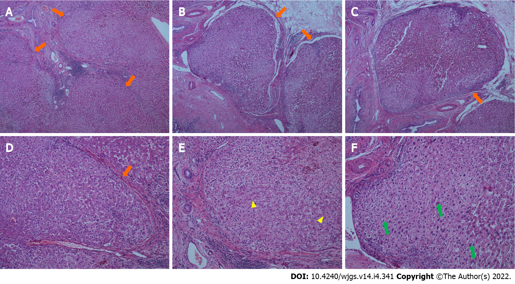
Figure 7 Histopathological findings in the resected liver (liver parenchyma).
A-C: Representative specimens showing proliferation of fibrous tissue in portal tracts dividing the liver parenchyma into irregular regenerative nodules (pseudolobules, orange arrows) that have lost the normal structure, and central veins (hematoxylin and eosin staining; 40 ×); D-F: Representative specimens showing pseudolobules (orange arrow) hepatic cords arranged irregularly, with foci of two layers of cells (yellow arrows), and large, occasionally binucleate cells (green arrows; hematoxylin and eosin staining; 100 ×).
- Citation: Fan WJ, Zou XJ. Subacute liver and respiratory failure after segmental hepatectomy for complicated hepatolithiasis with secondary biliary cirrhosis: A case report. World J Gastrointest Surg 2022; 14(4): 341-351
- URL: https://www.wjgnet.com/1948-9366/full/v14/i4/341.htm
- DOI: https://dx.doi.org/10.4240/wjgs.v14.i4.341


