Copyright
©The Author(s) 2022.
World J Gastrointest Surg. Mar 27, 2022; 14(3): 260-267
Published online Mar 27, 2022. doi: 10.4240/wjgs.v14.i3.260
Published online Mar 27, 2022. doi: 10.4240/wjgs.v14.i3.260
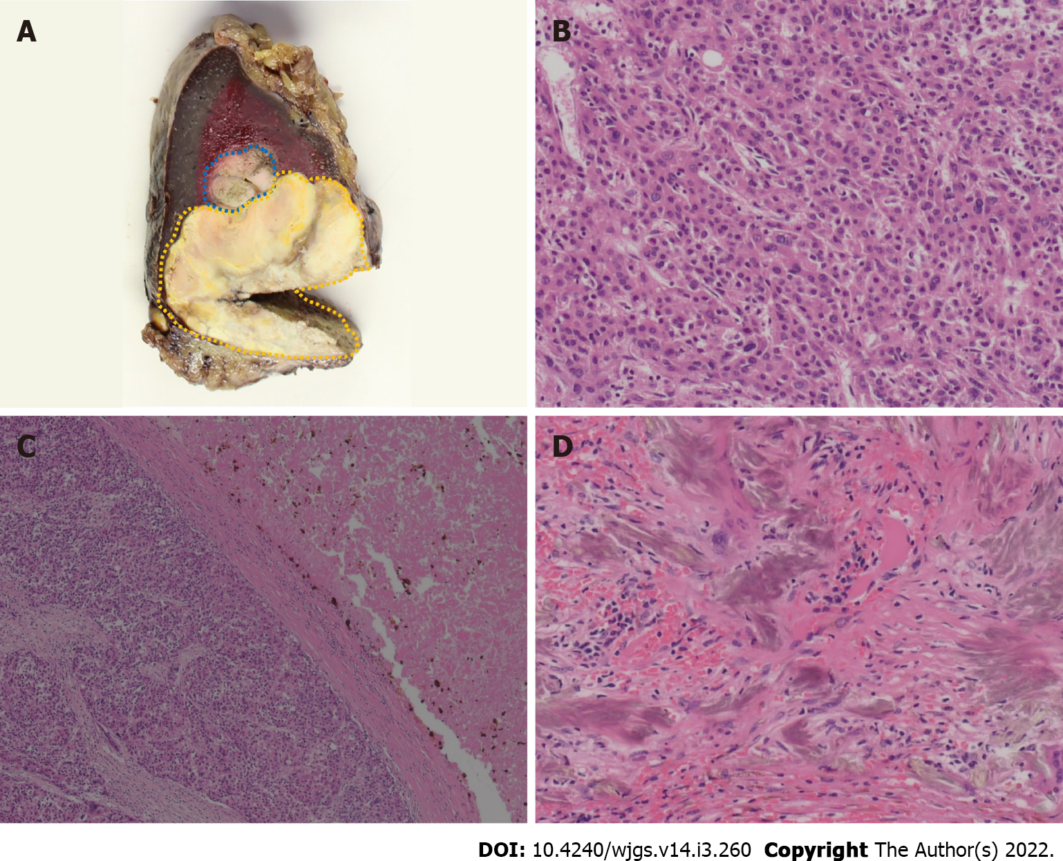
Figure 3 Pathological findings of metastatic splenic lesions.
A: Macroscopic finding shows that splenic lesions surrounded by fibrous capsule, and a border part (blue area) is distinguished from other parts (yellow area) with its color, suggesting viable lesions; B: Microscopic finding of viable tumor lesion shows moderately to poorly differentiated hepatocellular carcinoma. Hematoxylin-eosin stain, high-power field (× 200); C: Microscopic findings of mixed component of viable cells and necrotic tissue demonstrated that coagulative and partially liquefactive necrosis (right-side) is surrounded by fibrous capsule, and viable cells (left-side). Hematoxylin-eosin stain, low-power field (× 50); D: Gamma-Gandy bodies shown in the splenic lesions, suggesting previous history of portal hypertension due to portal vein tumor thrombosis. Hematoxylin-eosin stain, high-power field (× 200).
- Citation: Endo Y, Shimazu M, Sakuragawa T, Uchi Y, Edanami M, Sunamura K, Ozawa S, Chiba N, Kawachi S. Successful treatment with laparoscopic surgery and sequential multikinase inhibitor therapy for hepatocellular carcinoma: A case report. World J Gastrointest Surg 2022; 14(3): 260-267
- URL: https://www.wjgnet.com/1948-9366/full/v14/i3/260.htm
- DOI: https://dx.doi.org/10.4240/wjgs.v14.i3.260


