Copyright
©The Author(s) 2022.
World J Gastrointest Surg. Feb 27, 2022; 14(2): 200-210
Published online Feb 27, 2022. doi: 10.4240/wjgs.v14.i2.200
Published online Feb 27, 2022. doi: 10.4240/wjgs.v14.i2.200
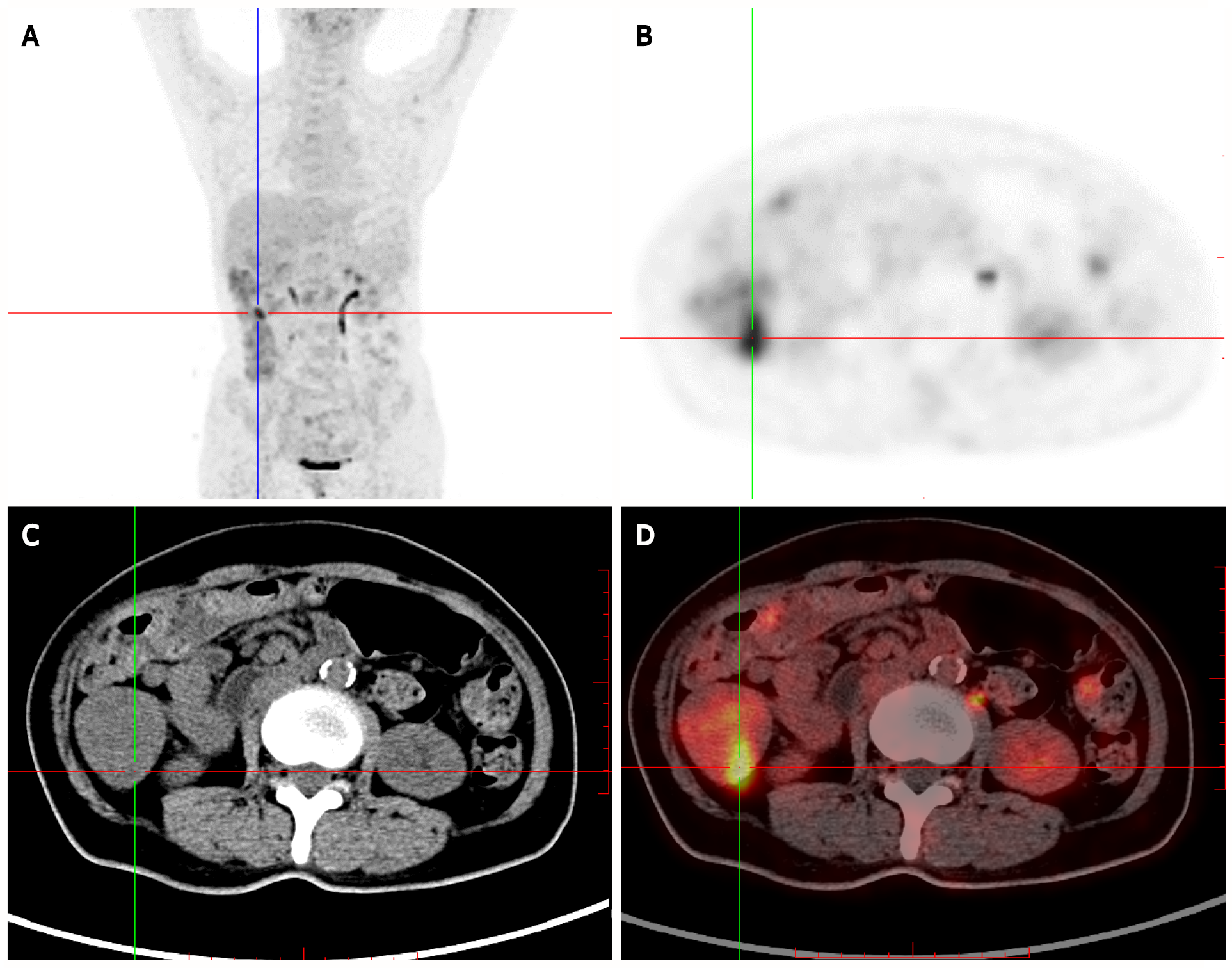
Figure 3 Positron-emission tomography/computed tomography showing multiple nodules with increased fluorodeoxyglucose uptake in the stomach wall, descending duodenum, and bulb, in the small intestine (obvious increase in the ileum), and the colon (obvious increase in the ascending colon).
Multiple nodular thickening with increased fluorodeoxyglucose (FDG) uptake was observed in the proximal rectum. A: Whole-body maximum intensity projection 18F-FDG and positron-emission tomography (PET) image; B: PET; C: Computed tomography (CT); D: PET/CT.
- Citation: Dong J, Ma TS, Tu JF, Chen YW. Surgery for Cronkhite-Canada syndrome complicated with intussusception: A case report and review of literature. World J Gastrointest Surg 2022; 14(2): 200-210
- URL: https://www.wjgnet.com/1948-9366/full/v14/i2/200.htm
- DOI: https://dx.doi.org/10.4240/wjgs.v14.i2.200


