Copyright
©The Author(s) 2022.
World J Gastrointest Surg. Dec 27, 2022; 14(12): 1425-1431
Published online Dec 27, 2022. doi: 10.4240/wjgs.v14.i12.1425
Published online Dec 27, 2022. doi: 10.4240/wjgs.v14.i12.1425
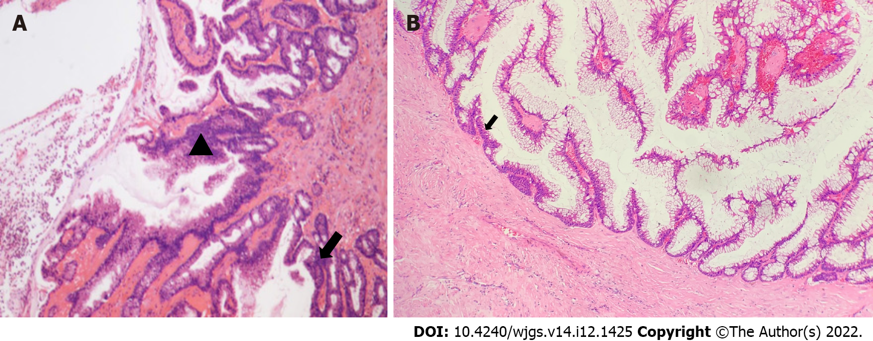
Figure 2 Histopathological analysis.
A: Moderate (▲)-to-severe (↑) dysplasia of the glandular epithelium; B: The columnar epithelial lining of the cyst wall had a large amount of mucus secretion, some of the epithelium had moderate dysplasia, and deranged smooth muscle bundles could be observed in the cyst wall (↑).
- Citation: Fang Y, Zhu Y, Liu WZ, Zhang XQ, Zhang Y, Wang K. Malignant transformation of perianal tailgut cyst: A case report. World J Gastrointest Surg 2022; 14(12): 1425-1431
- URL: https://www.wjgnet.com/1948-9366/full/v14/i12/1425.htm
- DOI: https://dx.doi.org/10.4240/wjgs.v14.i12.1425


