Copyright
©The Author(s) 2022.
World J Gastrointest Surg. Dec 27, 2022; 14(12): 1375-1386
Published online Dec 27, 2022. doi: 10.4240/wjgs.v14.i12.1375
Published online Dec 27, 2022. doi: 10.4240/wjgs.v14.i12.1375
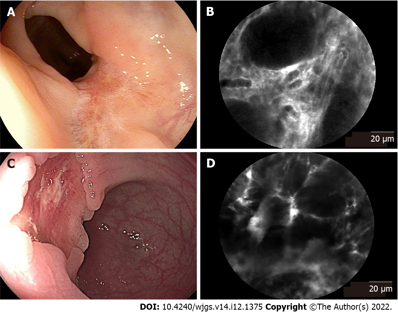
Figure 3 Endoscopic images and corresponding probe-based confocal laser endomicroscopy images of rectal tissues after neoadjuvant chemoradiotherapy.
A: Endoscopic image of rectal tissue with complete response, presenting a residual scar; B: Corresponding probe-based confocal laser endomicroscopy (pCLE) image showed regular crypts with thickening epithelium and enlarged vessels with fibrotic stroma; C: Endoscopic image of rectal tissue with partial response, presenting a residual tumor lesion; D: Corresponding pCLE image showed atypical glands with dark and irregular crypts and enlarged twisty vessels.
- Citation: Tan J, Ji HL, Hu YW, Li ZM, Zhuang BX, Deng HJ, Wang YN, Zheng JX, Jiang W, Yan J. Real-time in vivo distal margin selection using confocal laser endomicroscopy in transanal total mesorectal excision for rectal cancer. World J Gastrointest Surg 2022; 14(12): 1375-1386
- URL: https://www.wjgnet.com/1948-9366/full/v14/i12/1375.htm
- DOI: https://dx.doi.org/10.4240/wjgs.v14.i12.1375


