Copyright
©The Author(s) 2022.
World J Gastrointest Surg. Dec 27, 2022; 14(12): 1375-1386
Published online Dec 27, 2022. doi: 10.4240/wjgs.v14.i12.1375
Published online Dec 27, 2022. doi: 10.4240/wjgs.v14.i12.1375
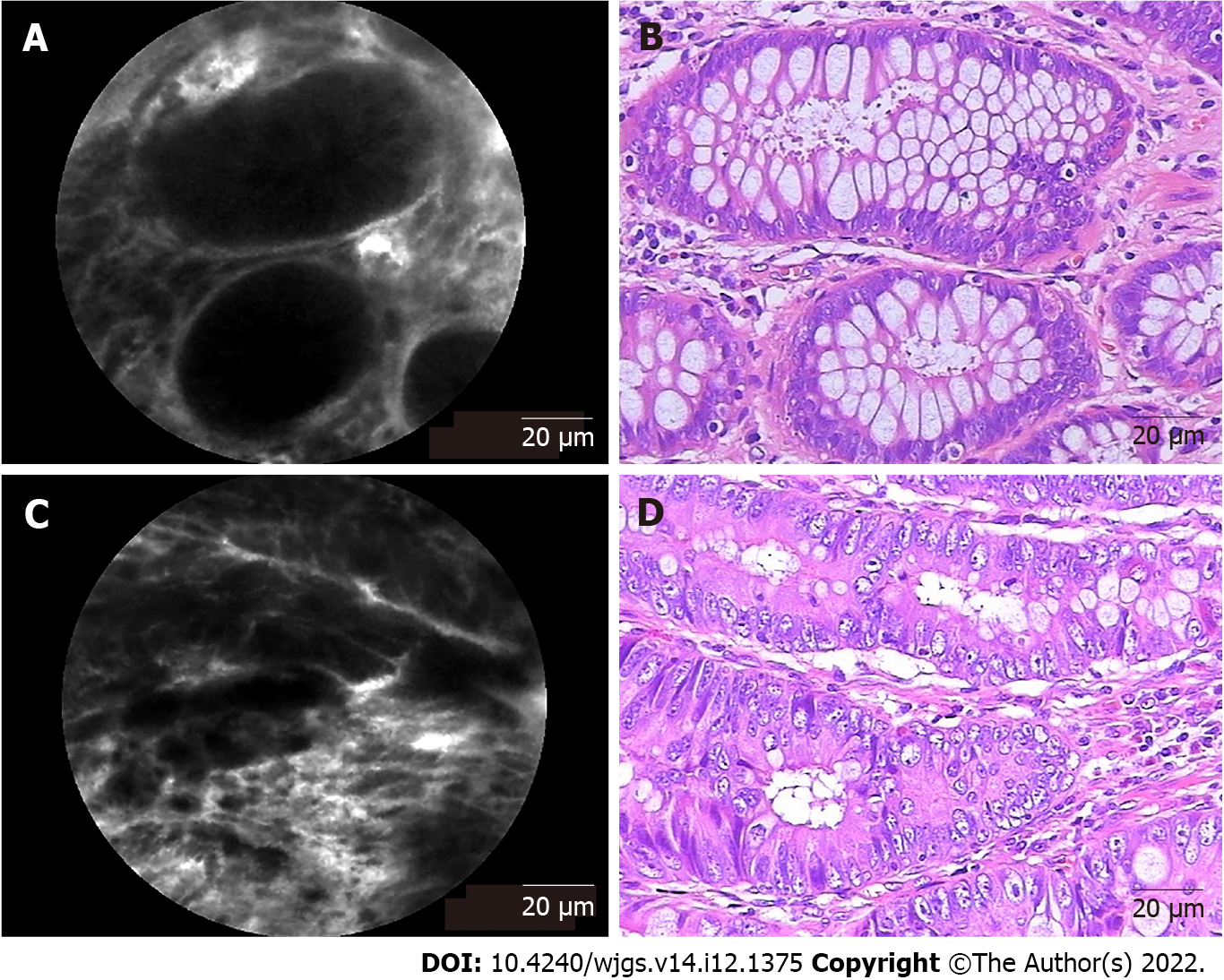
Figure 2 Representative probe-based confocal laser endomicroscopy images and corresponding hematoxylin and eosin-stained images of rectal tissues.
A: Probe-based confocal laser endomicroscopy (pCLE) image of normal tissue presenting normal round crypt structures with regular luminal openings covered by a homogeneous single-cell-layered epithelium with dark goblet cells and regular narrow vessels with hexagonal, honeycomb appearance surrounding crypts; B: Corresponding image of normal rectal tissue stained by hematoxylin and eosin (H&E); C: pCLE image of rectal neoplastic tissues manifesting as dark and irregularly thickened epithelium with decreased volume of lamina propria and dilated, distorted vessels with elevated leakage. The epithelium was dark and irregularly thickened, and the vessels were dilated; D: Corresponding image of H&E-stained rectal adenocarcinoma tissue.
- Citation: Tan J, Ji HL, Hu YW, Li ZM, Zhuang BX, Deng HJ, Wang YN, Zheng JX, Jiang W, Yan J. Real-time in vivo distal margin selection using confocal laser endomicroscopy in transanal total mesorectal excision for rectal cancer. World J Gastrointest Surg 2022; 14(12): 1375-1386
- URL: https://www.wjgnet.com/1948-9366/full/v14/i12/1375.htm
- DOI: https://dx.doi.org/10.4240/wjgs.v14.i12.1375


