Copyright
©The Author(s) 2022.
World J Gastrointest Surg. Dec 27, 2022; 14(12): 1375-1386
Published online Dec 27, 2022. doi: 10.4240/wjgs.v14.i12.1375
Published online Dec 27, 2022. doi: 10.4240/wjgs.v14.i12.1375
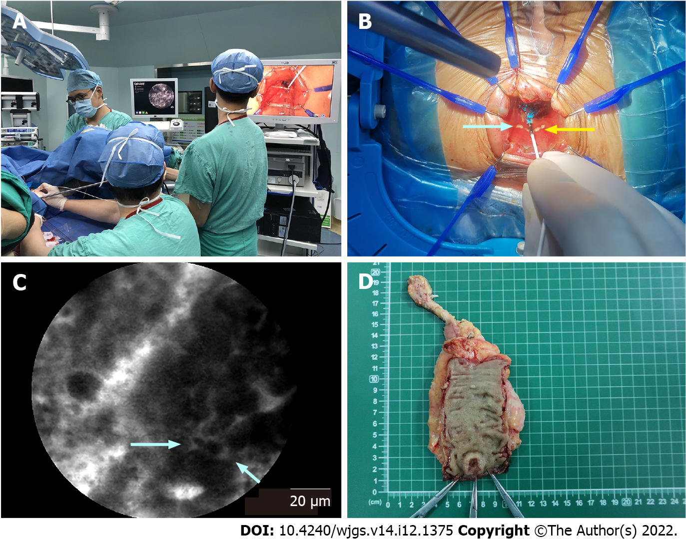
Figure 1 Selection of the distal resection margin guided by probe-based confocal laser endomicroscopy.
A: During transanal total mesorectal excision, the surgeon used the tip of the probe in direct contact with the tissues. In the meantime, the pathologist analyzed the real-time probe-based confocal laser endomicroscopy (pCLE) videos; B: A dot (white arrow) was marked by an electric scalpel at the distal edge of the tumor, which was determined by pCLE optical biopsy. Then, the distal resection margin (DRM) (yellow arrow) was marked 5-10 mm below the marked dot; C: In pCLE videos, the distal edge of the tumor (white arrow) was determined at the end of the distorted structures (dark, irregularly thickened epithelium); D: Conventional samples were collected for histology at the marked dot and DRM after surgery.
- Citation: Tan J, Ji HL, Hu YW, Li ZM, Zhuang BX, Deng HJ, Wang YN, Zheng JX, Jiang W, Yan J. Real-time in vivo distal margin selection using confocal laser endomicroscopy in transanal total mesorectal excision for rectal cancer. World J Gastrointest Surg 2022; 14(12): 1375-1386
- URL: https://www.wjgnet.com/1948-9366/full/v14/i12/1375.htm
- DOI: https://dx.doi.org/10.4240/wjgs.v14.i12.1375


