Copyright
©The Author(s) 2022.
World J Gastrointest Surg. Dec 27, 2022; 14(12): 1329-1339
Published online Dec 27, 2022. doi: 10.4240/wjgs.v14.i12.1329
Published online Dec 27, 2022. doi: 10.4240/wjgs.v14.i12.1329
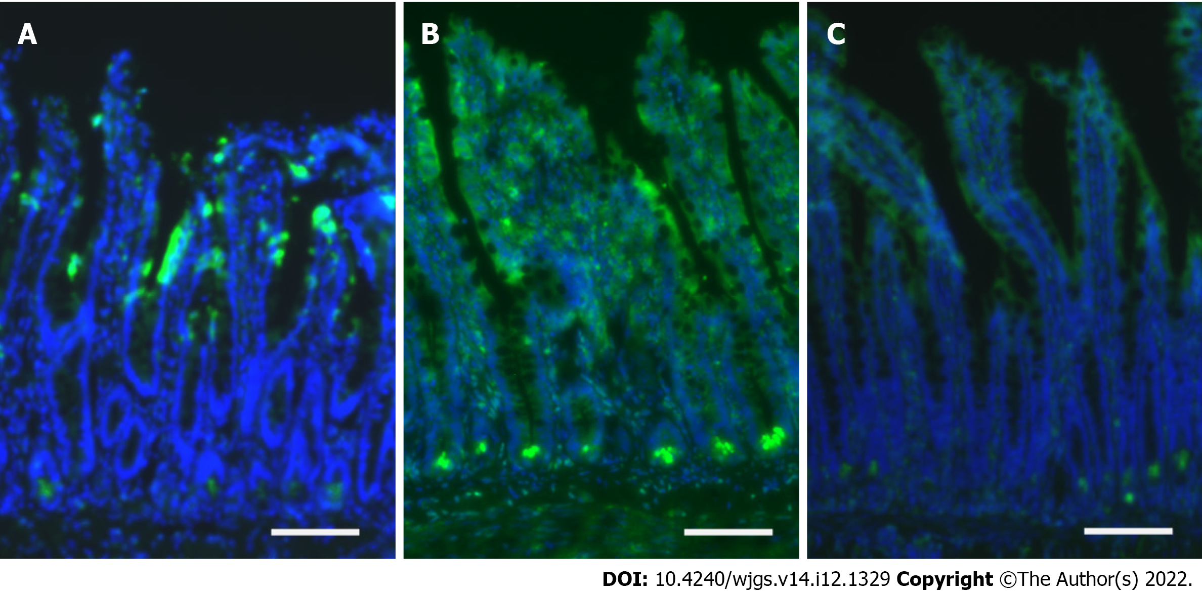
Figure 2 Intestinal stem cell injury by ischemia-reperfusion injury.
A: Immunofluorescence staining with caspase-3 (green) revealed mucosal injury at the epithelial layer closed to the tip of the villi in the ischemia group; B: Extensive apoptosis was identified at the crypt base and the whole epithelial layer in the reperfusion group; C: Apoptosis at the crypt base was limited in the hydrogen group. Scale bar: 100 μm.
- Citation: Yamamoto R, Suzuki S, Homma K, Yamaguchi S, Sujino T, Sasaki J. Hydrogen gas and preservation of intestinal stem cells in mesenteric ischemia and reperfusion. World J Gastrointest Surg 2022; 14(12): 1329-1339
- URL: https://www.wjgnet.com/1948-9366/full/v14/i12/1329.htm
- DOI: https://dx.doi.org/10.4240/wjgs.v14.i12.1329


