Copyright
©The Author(s) 2022.
World J Gastrointest Surg. Oct 27, 2022; 14(10): 1169-1178
Published online Oct 27, 2022. doi: 10.4240/wjgs.v14.i10.1169
Published online Oct 27, 2022. doi: 10.4240/wjgs.v14.i10.1169
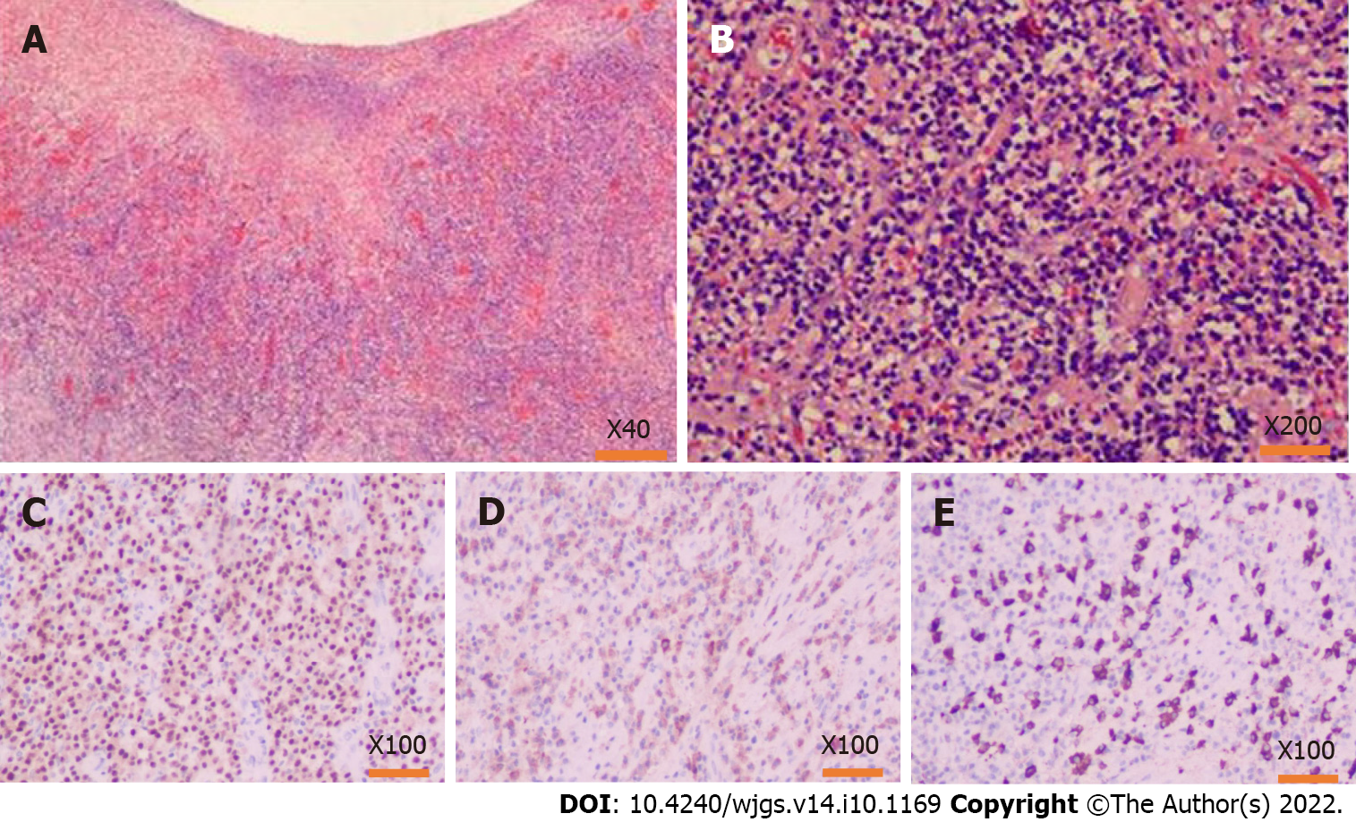
Figure 7 Pathology findings after the operation.
A and B: Hematoxylin-eosin staining of the lesion tissues, inflammatory cell infiltration with plasma cells and neutrophils and increased immunoglobulin G4 (IgG4) positive cells could be seen in the resection colonic mass, accompanied by tissue fibrosis and obliterative phlebitis; C: MMU1 staining showed a high expression state, suggesting a large number of plasma cells infiltration; D: Immunohistochemistry showed a lot of positive IgG staining in the lesion area; E: IgG4 also showed much positive staining in the lesion area, and the number of IgG4-positive cells in the main core area was as high as 110/HPF.
- Citation: Zhan WL, Liu L, Jiang W, He FX, Qu HT, Cao ZX, Xu XS. Immunoglobulin G4-related disease in the sigmoid colon in patient with severe colonic fibrosis and obstruction: A case report. World J Gastrointest Surg 2022; 14(10): 1169-1178
- URL: https://www.wjgnet.com/1948-9366/full/v14/i10/1169.htm
- DOI: https://dx.doi.org/10.4240/wjgs.v14.i10.1169


