Copyright
©The Author(s) 2022.
World J Gastrointest Surg. Oct 27, 2022; 14(10): 1161-1168
Published online Oct 27, 2022. doi: 10.4240/wjgs.v14.i10.1161
Published online Oct 27, 2022. doi: 10.4240/wjgs.v14.i10.1161
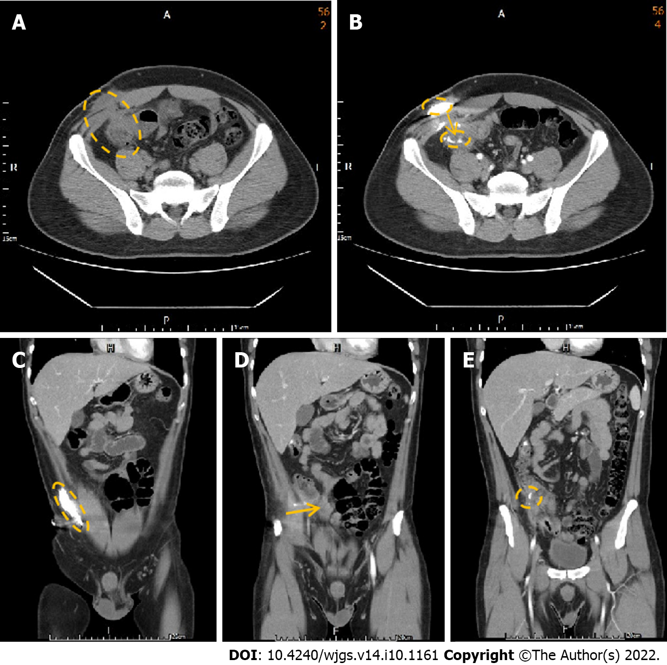
Figure 2 Abdominal computed tomography with contrast injection from the abdominal opening.
A: The axial computed tomography (CT) demonstrates the colon in close contact with the abdominal wall; B: The axial CT with contrast injection into the abdominal carbuncle demonstrates the canal between the abdominal wall and colon; C: Contrast was injected from the carbuncle and accumulated in the subcutaneous area, indicating abscess formation; D: Contrast dye extended through the canal between the abdominal wall and colon; E: Contrast finally arrived at the colon.
- Citation: Wu TY, Lo KH, Chen CY, Hu JM, Kang JC, Pu TW. Cecocutaneous fistula diagnosed by computed tomography fistulography: A case report. World J Gastrointest Surg 2022; 14(10): 1161-1168
- URL: https://www.wjgnet.com/1948-9366/full/v14/i10/1161.htm
- DOI: https://dx.doi.org/10.4240/wjgs.v14.i10.1161


