Copyright
©The Author(s) 2022.
World J Gastrointest Surg. Oct 27, 2022; 14(10): 1141-1149
Published online Oct 27, 2022. doi: 10.4240/wjgs.v14.i10.1141
Published online Oct 27, 2022. doi: 10.4240/wjgs.v14.i10.1141
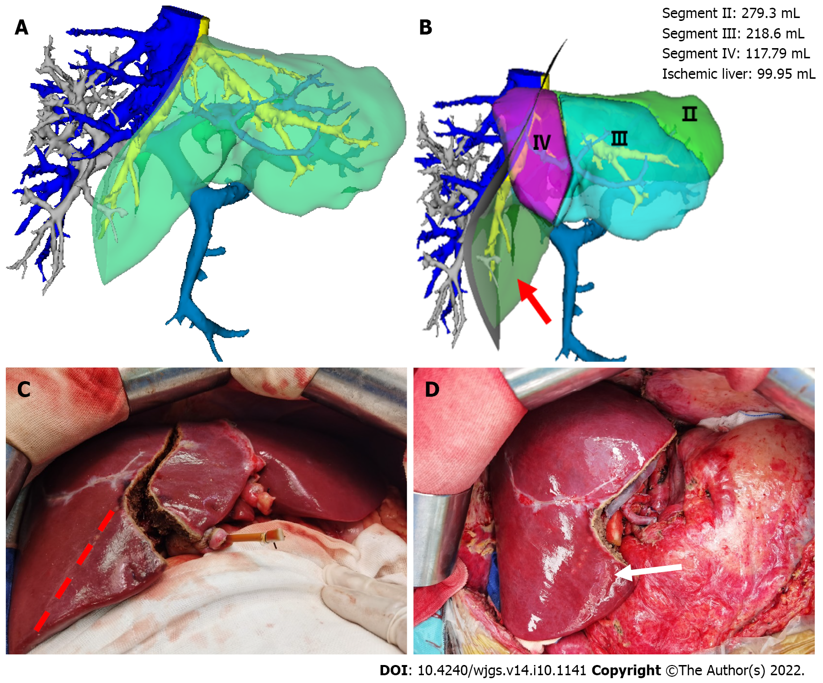
Figure 3 Left and right half-split liver transplantation was performed according to the portal vein blood flow topology liver segmentation method.
There was no ischemic change in the liver segment after reflow. A: Left and right half liver splitting were simulated based on the Couinaud liver segmentation method, while the middle hepatic vein was split in the middle; B: The simulation of left and right half liver splitting based on the portal vein blood flow topology liver segmentation method. It can be seen that a portion of segment V liver tissues (99.95 mL) is partitioned into the left liver (arrow); C: Surgery based on the portal vein blood flow topology liver segmentation method was implemented for left and right half liver splitting instead of the Couinaud liver segmentation method (red dotted line); D: No ischemic changes in the hepatic segment after reflow (white arrow).
- Citation: Zhao D, Zhang KJ, Fang TS, Yan X, Jin X, Liang ZM, Tang JX, Xie LJ. Topological approach of liver segmentation based on 3D visualization technology in surgical planning for split liver transplantation. World J Gastrointest Surg 2022; 14(10): 1141-1149
- URL: https://www.wjgnet.com/1948-9366/full/v14/i10/1141.htm
- DOI: https://dx.doi.org/10.4240/wjgs.v14.i10.1141


