Copyright
©The Author(s) 2021.
World J Gastrointest Surg. May 27, 2021; 13(5): 507-515
Published online May 27, 2021. doi: 10.4240/wjgs.v13.i5.507
Published online May 27, 2021. doi: 10.4240/wjgs.v13.i5.507
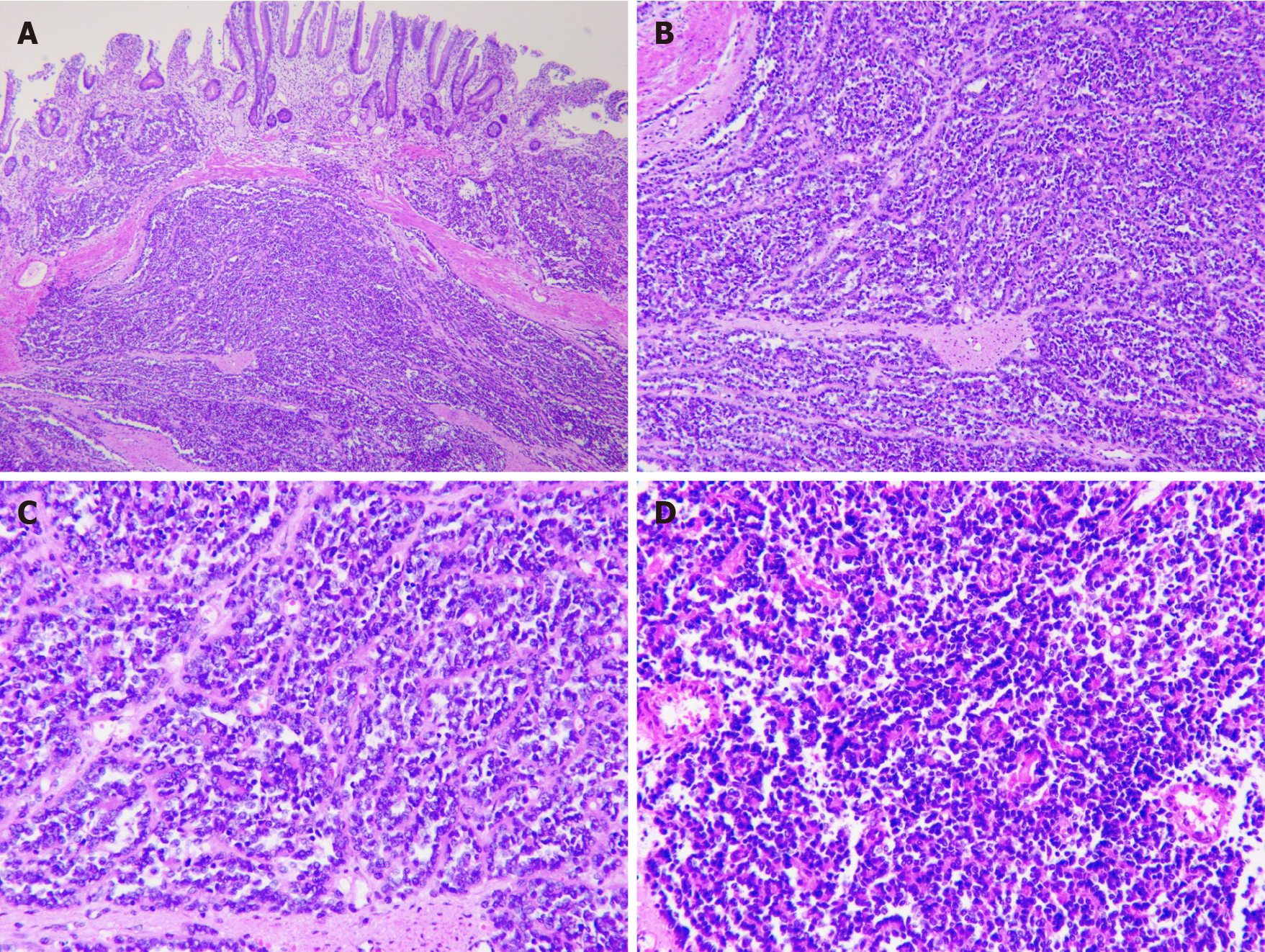
Figure 2 Immunohistochemical analysis.
A: Low magnification of the resected sample using formalin-fixed (magnification: × 40); B: Paraffin-embedded sections of tumor stained with hematoxylin and eosin demonstrating sheets of small (magnification: × 100); C: Round-to-spindle, uniform tumor cells with clear cytoplasm (magnification: × 200); D: Higher magnification of C (magnification: × 200).
- Citation: Shadhu K, Ramlagun-Mungur D, Ping XC. Ewing sarcoma of the jejunum: A case report and literature review . World J Gastrointest Surg 2021; 13(5): 507-515
- URL: https://www.wjgnet.com/1948-9366/full/v13/i5/507.htm
- DOI: https://dx.doi.org/10.4240/wjgs.v13.i5.507


