Copyright
©The Author(s) 2021.
World J Gastrointest Surg. May 27, 2021; 13(5): 406-418
Published online May 27, 2021. doi: 10.4240/wjgs.v13.i5.406
Published online May 27, 2021. doi: 10.4240/wjgs.v13.i5.406
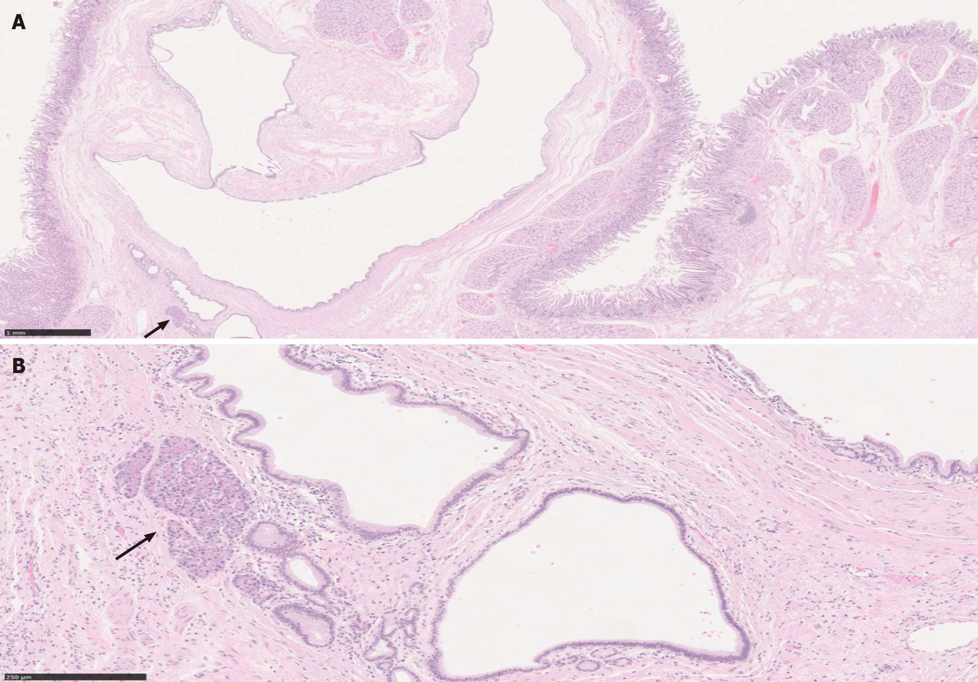
Figure 4 Paraduodenal pancreatitis.
A: This image shows cyst formation, Brunner gland hyperplasia, and ectopic pancreatic tissue (solid arrow) with associated inflammation, consistent with paraduodenal pancreatitis, in a patient with history of significant alcohol use [hematoxylin & eosin (H&E), 20 ×, scale 1 mm]; B: A higher magnification to show the ectopic pancreatic tissue in association with other elements (H&E, 130 ×, scale 250 µm).
- Citation: Aldyab M, El Jabbour T, Parilla M, Lee H. Benign vs malignant pancreatic lesions: Molecular insights to an ongoing debate. World J Gastrointest Surg 2021; 13(5): 406-418
- URL: https://www.wjgnet.com/1948-9366/full/v13/i5/406.htm
- DOI: https://dx.doi.org/10.4240/wjgs.v13.i5.406


