Copyright
©The Author(s) 2021.
World J Gastrointest Surg. Apr 27, 2021; 13(4): 340-354
Published online Apr 27, 2021. doi: 10.4240/wjgs.v13.i4.340
Published online Apr 27, 2021. doi: 10.4240/wjgs.v13.i4.340
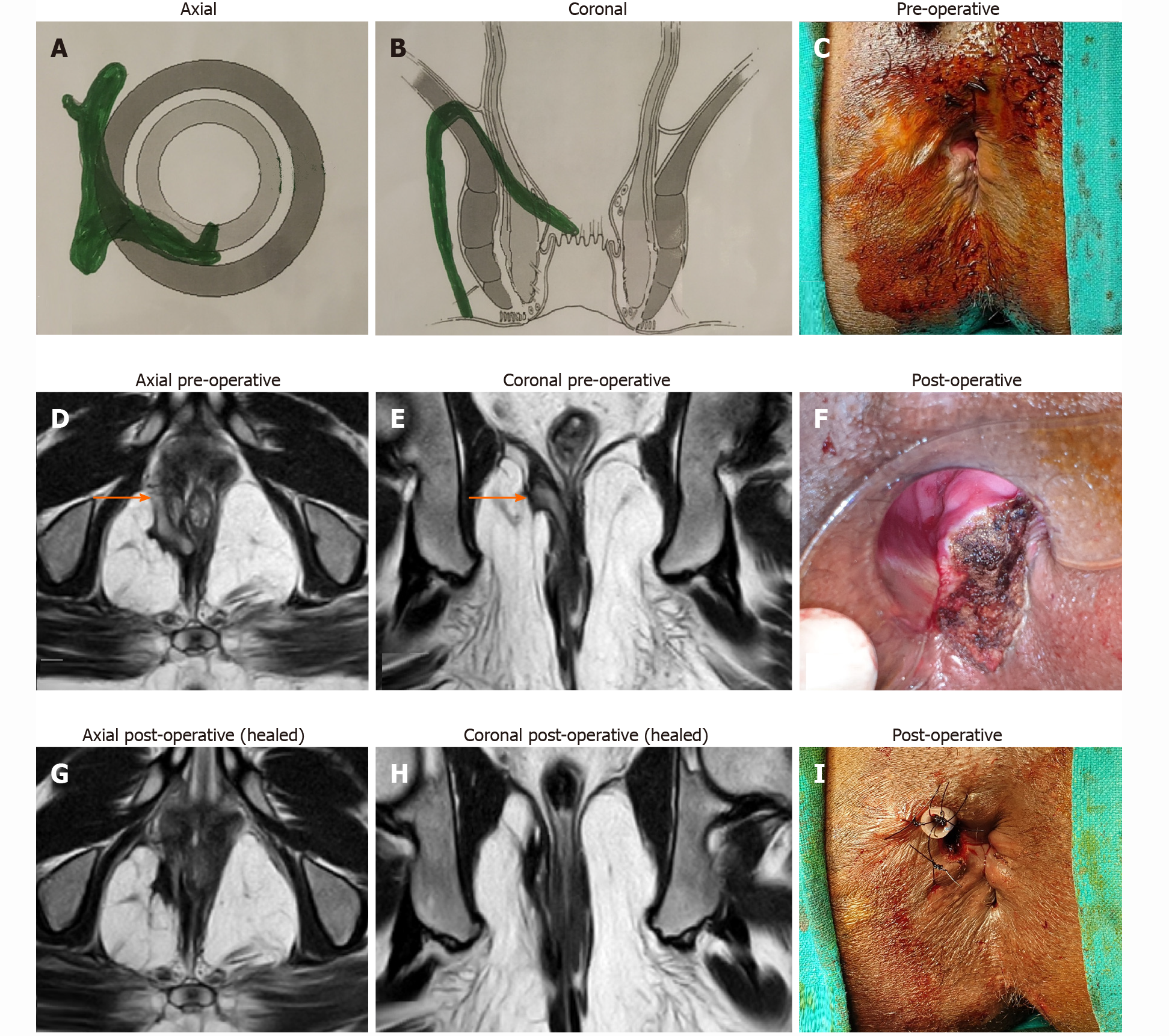
Figure 3 A 35-year-old male patient with a suprasphincteric anal fistula managed by transanal opening of intersphincteric spaceprocedure.
A: Axial section (Schematic diagram); B: Coronal section (Schematic diagram); C: Preoperative photograph; D: Preoperative T2-weighted magnetic resonance image (MRI) axial section; E: T2-weighted preoperative MRI coronal section; F: Postoperative photograph showing the transanal opening of intersphincteric space wound, the laid open intersphincteric portion of the fistula tract, in the anal canal; G: Postoperative T2-weighted MRI axial section 3 mo after surgery showing healed fistula tracts; H: Postoperative T2-weighted MRI coronal section 3 mo after surgery showing healed fistula tracts; I: Postoperative photograph showing the final picture and a tube inserted in the tract in right ischiorectal fossa. The tube was sutured to the skin with monofilament non-absorbable 2-0 nylon. Orange arrows show fistula tracts.
- Citation: Garg P, Kaur B, Goyal A, Yagnik VD, Dawka S, Menon GR. Lessons learned from an audit of 1250 anal fistula patients operated at a single center: A retrospective review. World J Gastrointest Surg 2021; 13(4): 340-354
- URL: https://www.wjgnet.com/1948-9366/full/v13/i4/340.htm
- DOI: https://dx.doi.org/10.4240/wjgs.v13.i4.340


