Copyright
©The Author(s) 2021.
World J Gastrointest Surg. Mar 27, 2021; 13(3): 323-329
Published online Mar 27, 2021. doi: 10.4240/wjgs.v13.i3.323
Published online Mar 27, 2021. doi: 10.4240/wjgs.v13.i3.323
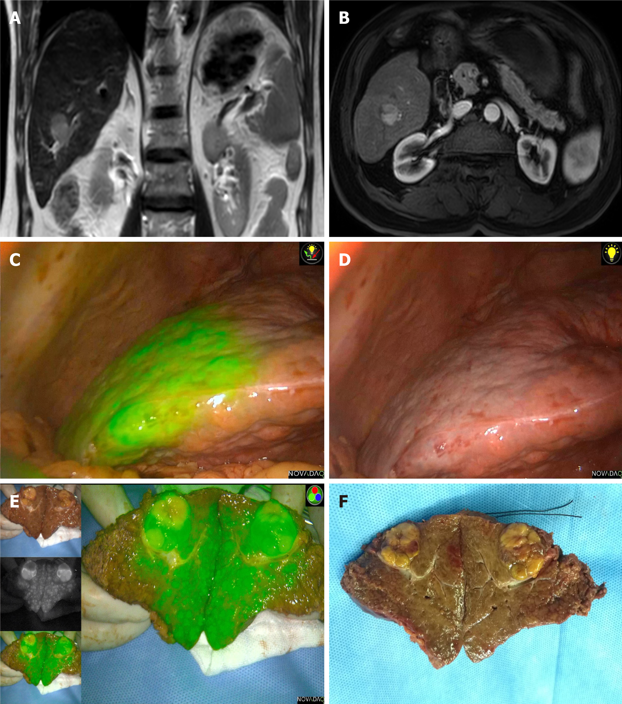
Figure 3 Imaging examinations of Case 3.
A and B: Preoperative magnetic resonance imaging examination indicated a hepatocellular carcinoma approximately 20 mm in diameter located at segment VI; C: Fluorescent staining of segment VI; D: The ischemic line on the liver surface after clipping the segment VI pedicle; E and F: Postoperative specimen confirmed the Glissonian pedicle oppressed by the tumor and the stained segment.
- Citation: Han HW, Shi N, Zou YP, Zhang YP, Lin Y, Yin Z, Jian ZX, Jin HS. Functional anatomical hepatectomy guided by indocyanine green fluorescence imaging in patients with localized cholestasis: Report of four cases. World J Gastrointest Surg 2021; 13(3): 323-329
- URL: https://www.wjgnet.com/1948-9366/full/v13/i3/323.htm
- DOI: https://dx.doi.org/10.4240/wjgs.v13.i3.323


