Copyright
©The Author(s) 2021.
World J Gastrointest Surg. Nov 27, 2021; 13(11): 1315-1326
Published online Nov 27, 2021. doi: 10.4240/wjgs.v13.i11.1315
Published online Nov 27, 2021. doi: 10.4240/wjgs.v13.i11.1315
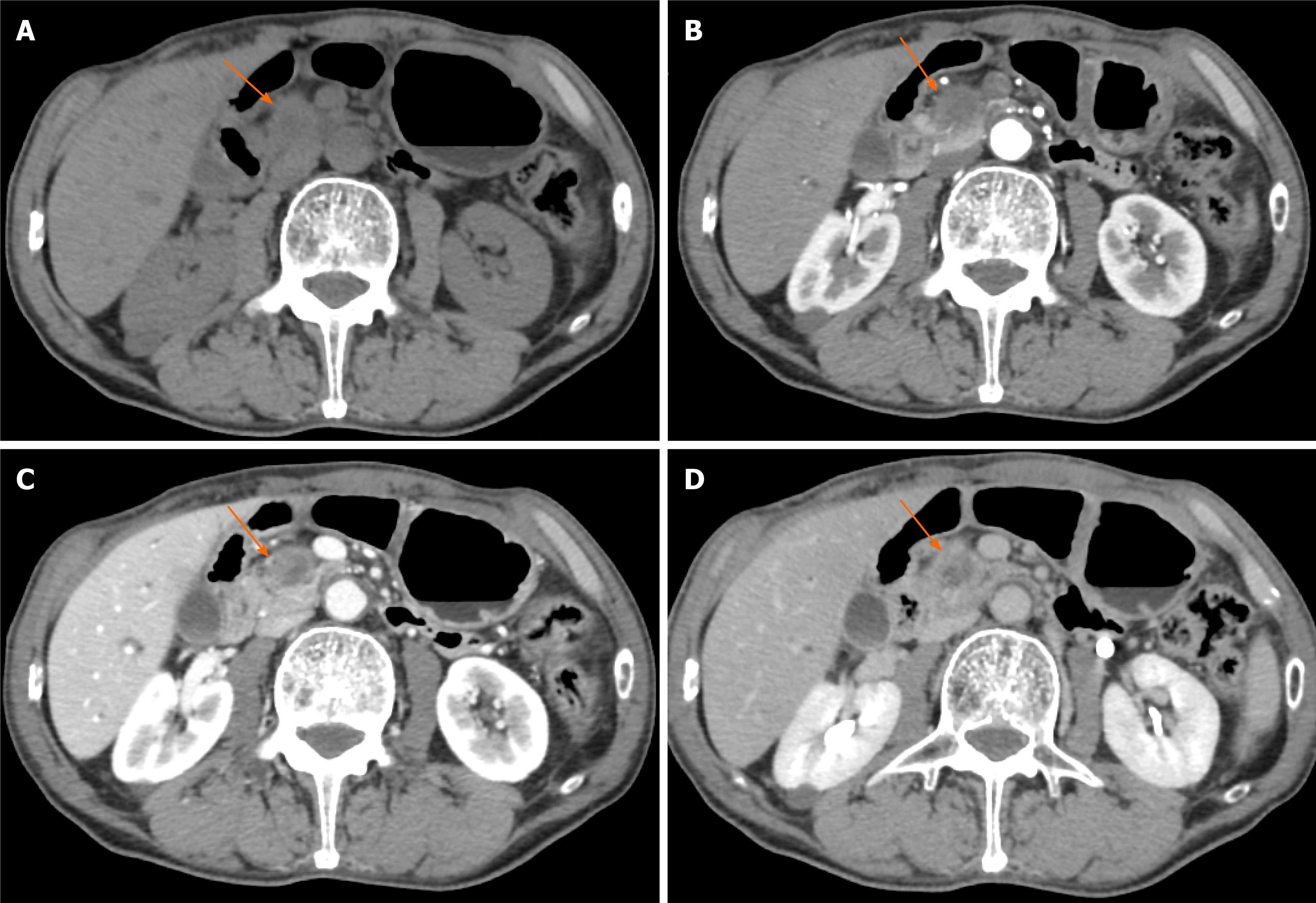
Figure 3 Typical CT features of pancreatic head carcinoma in a 73-year-old male patient.
A: On noncontrast CT imaging a slightly low-density mass (arrow) in the pancreatic head area was identified; B–D: On contrast CT images, the tumor shows an avascular tumor with a lower density than normal pancreatic parenchyma on arterial phase (B), venous phase (C), and delay phase (D). CT: Computed tomography.
- Citation: Feng P, Cheng B, Wang ZD, Liu JG, Fan W, Liu H, Qi CY, Pan JJ. Application and progress of medical imaging in total mesopancreas excision for pancreatic head carcinoma. World J Gastrointest Surg 2021; 13(11): 1315-1326
- URL: https://www.wjgnet.com/1948-9366/full/v13/i11/1315.htm
- DOI: https://dx.doi.org/10.4240/wjgs.v13.i11.1315


