Copyright
©The Author(s) 2021.
World J Gastrointest Surg. Nov 27, 2021; 13(11): 1315-1326
Published online Nov 27, 2021. doi: 10.4240/wjgs.v13.i11.1315
Published online Nov 27, 2021. doi: 10.4240/wjgs.v13.i11.1315
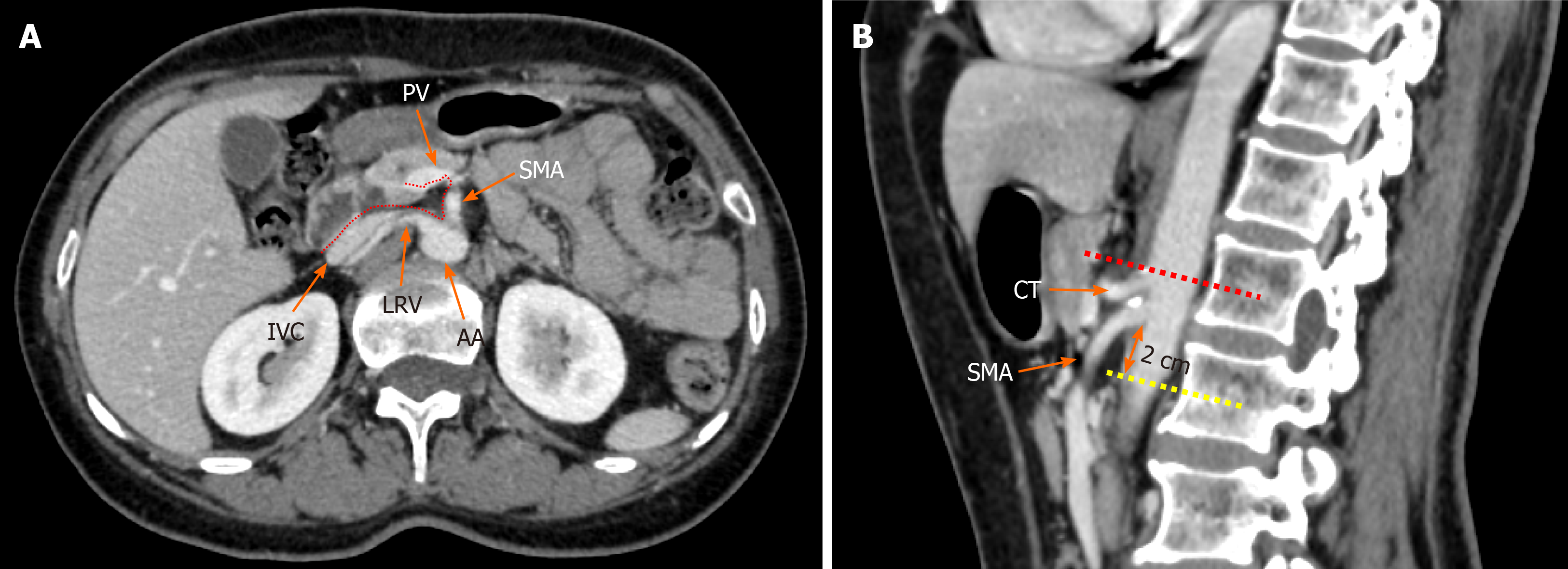
Figure 1 Radiological depiction of the mesopancreas in computed tomography.
A: The dotted line outlines the boundary of the mesopancreas, a region identified as the retro pancreatic retro portal tissue; B: The inferior boundary of the mesopancreas is 2 cm below the origin of superior mesenteric artery. PV: Portal vein; SMA: Superior mesenteric artery; LRV: Left renal vein; IVC: Inferior vena cava; AA: Aorta artery; CT: Celiac trunk.
- Citation: Feng P, Cheng B, Wang ZD, Liu JG, Fan W, Liu H, Qi CY, Pan JJ. Application and progress of medical imaging in total mesopancreas excision for pancreatic head carcinoma. World J Gastrointest Surg 2021; 13(11): 1315-1326
- URL: https://www.wjgnet.com/1948-9366/full/v13/i11/1315.htm
- DOI: https://dx.doi.org/10.4240/wjgs.v13.i11.1315


