Copyright
©The Author(s) 2021.
World J Gastrointest Surg. Oct 27, 2021; 13(10): 1285-1292
Published online Oct 27, 2021. doi: 10.4240/wjgs.v13.i10.1285
Published online Oct 27, 2021. doi: 10.4240/wjgs.v13.i10.1285
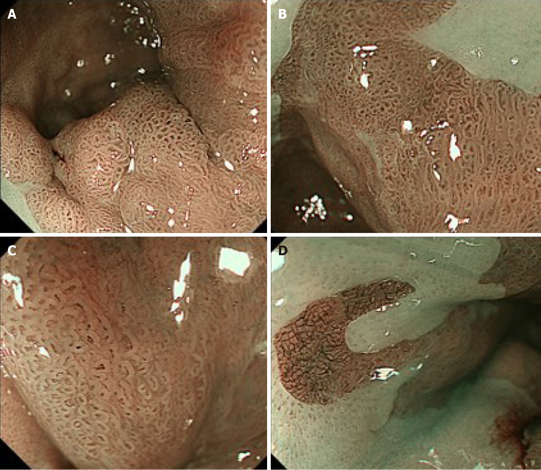
Figure 2 Images of narrow-band imaging magnification endoscopy.
A: Narrow-band imaging magnification endoscopy (NBI-M) shows a visible mucosal pattern with villous structures in protruded or elevated portions, as well as in the surrounding flat portions in the long-segment Barrett’s esophagus (LSBE); B: The villous patterns were rated as irregular because they showed variety in size and existed in a high density; C: In most of surrounding areas of the LSBE, mucosal patterns showed an irregular villous pattern [similar to the image (C)], and vascular patterns were rated as irregular because they showed a variety of forms and calibers under NBI-M observation with high magnification; D: In several flat areas, NBI-M demonstrated an invisible mucosal pattern with an irregular vascular pattern forming a network-like structure with a variety in caliber.
- Citation: Abe K, Goda K, Kanamori A, Suzuki T, Yamamiya A, Takimoto Y, Arisaka T, Hoshi K, Sugaya T, Majima Y, Tominaga K, Iijima M, Hirooka S, Yamagishi H, Irisawa A. Whole circumferential endoscopic submucosal dissection of superficial adenocarcinoma in long-segment Barrett's esophagus: A case report. World J Gastrointest Surg 2021; 13(10): 1285-1292
- URL: https://www.wjgnet.com/1948-9366/full/v13/i10/1285.htm
- DOI: https://dx.doi.org/10.4240/wjgs.v13.i10.1285


