Copyright
©The Author(s) 2021.
World J Gastrointest Surg. Oct 27, 2021; 13(10): 1285-1292
Published online Oct 27, 2021. doi: 10.4240/wjgs.v13.i10.1285
Published online Oct 27, 2021. doi: 10.4240/wjgs.v13.i10.1285
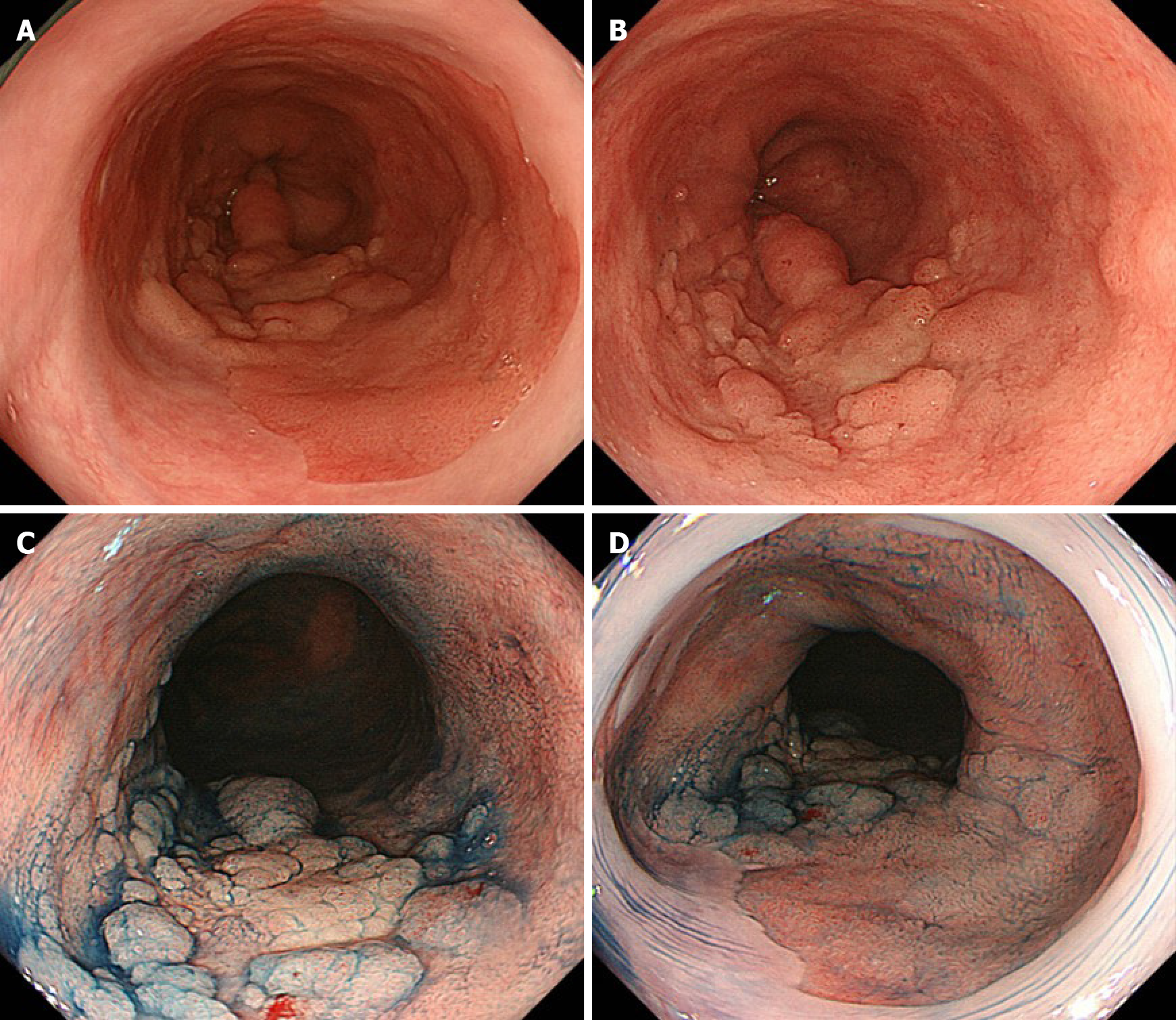
Figure 1 Images of esophagogastroduodenoscopy.
A and B: Conventional white-light endoscopy shows a nodular aggregated protruded lesion in the long-segment Barrett’s esophagus (C 3.5 M 4); C and D: Chromoendoscopy with indigo carmine shows the protruded lesion with irregularly sized granules. No obvious lesion was found in the mucosal surface of Barrett's esophagus other than the nodular aggregated protruded lesion.
- Citation: Abe K, Goda K, Kanamori A, Suzuki T, Yamamiya A, Takimoto Y, Arisaka T, Hoshi K, Sugaya T, Majima Y, Tominaga K, Iijima M, Hirooka S, Yamagishi H, Irisawa A. Whole circumferential endoscopic submucosal dissection of superficial adenocarcinoma in long-segment Barrett's esophagus: A case report. World J Gastrointest Surg 2021; 13(10): 1285-1292
- URL: https://www.wjgnet.com/1948-9366/full/v13/i10/1285.htm
- DOI: https://dx.doi.org/10.4240/wjgs.v13.i10.1285


