Copyright
©The Author(s) 2021.
World J Gastrointest Surg. Jan 27, 2021; 13(1): 30-49
Published online Jan 27, 2021. doi: 10.4240/wjgs.v13.i1.30
Published online Jan 27, 2021. doi: 10.4240/wjgs.v13.i1.30
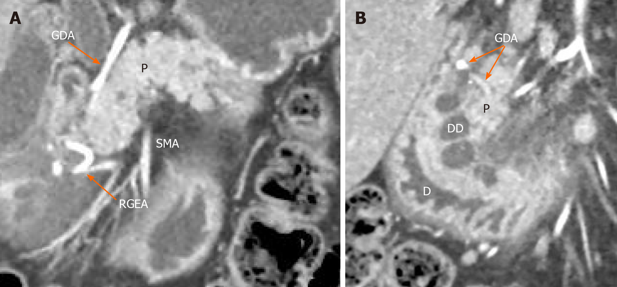
Figure 2 Isolated form of cystic dystrophy of the duodenal wall.
Arterial phase. Coronal view. A: Deformation and thickening of the medial wall of the duodenum (D), major papilla surrounded by well-defined cysts located in the submucosa (DD). The gastroduodenal artery is shifted forward and to the left, lying in the groove between the unaffected pancreatic head (P) and duodenal wall; B: Unchanged orthotopic pancreas. Only the duodenum and the groove are involved. SMA: Superior mesenteric artery; GDA: Gastroduodenal artery; RGEA: Right gastro-epiploic artery.
- Citation: Egorov V, Petrov R, Schegolev A, Dubova E, Vankovich A, Kondratyev E, Dobriakov A, Kalinin D, Schvetz N, Poputchikova E. Pancreas-preserving duodenal resections vs pancreatoduodenectomy for groove pancreatitis. Should we revisit treatment algorithm for groove pancreatitis? World J Gastrointest Surg 2021; 13(1): 30-49
- URL: https://www.wjgnet.com/1948-9366/full/v13/i1/30.htm
- DOI: https://dx.doi.org/10.4240/wjgs.v13.i1.30


