Copyright
©The Author(s) 2020.
World J Gastrointest Surg. Jun 27, 2020; 12(6): 298-306
Published online Jun 27, 2020. doi: 10.4240/wjgs.v12.i6.298
Published online Jun 27, 2020. doi: 10.4240/wjgs.v12.i6.298
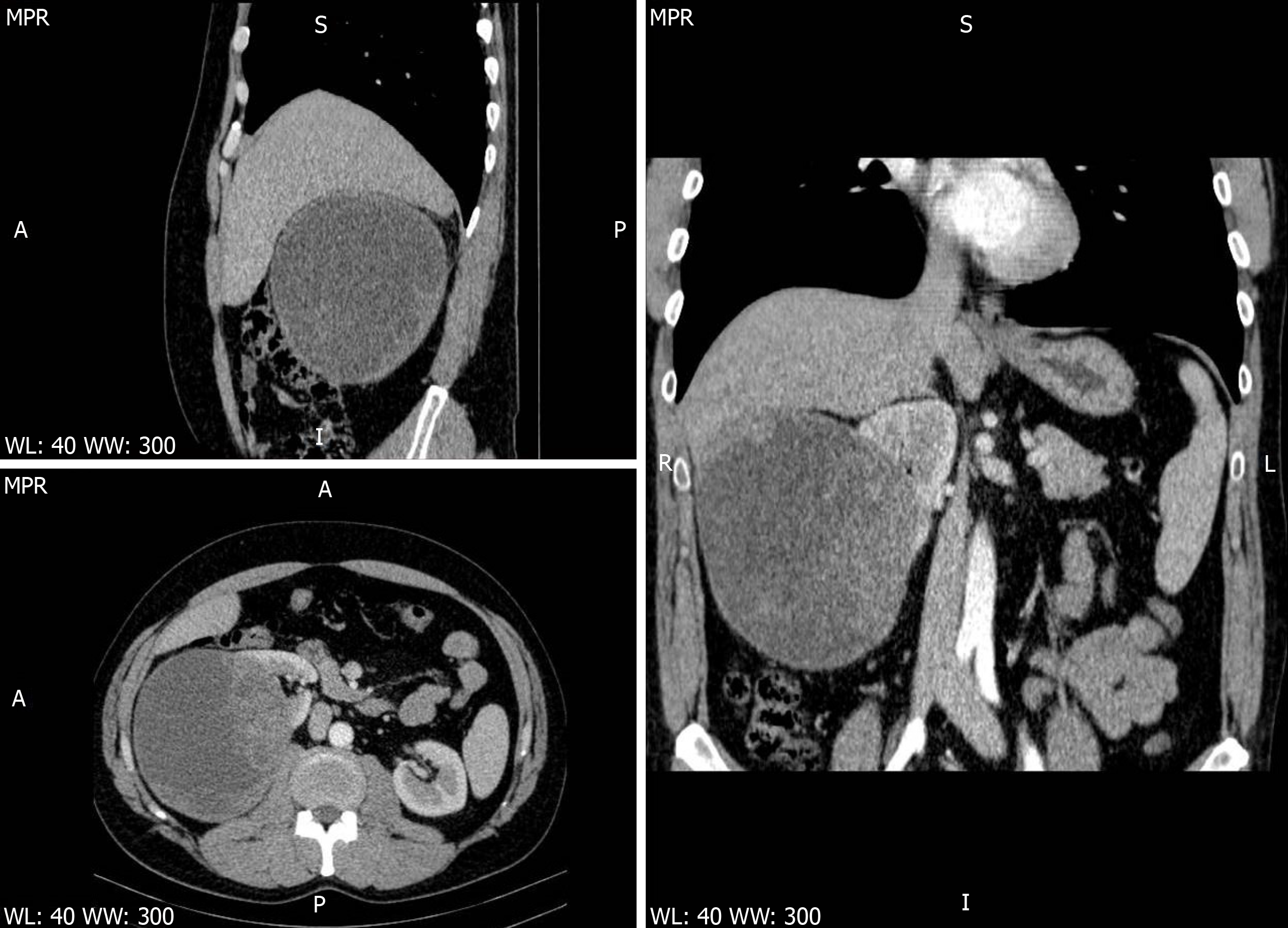
Figure 1 Computed tomography images.
Contrast-enhanced abdominal computed tomography scan thin multiplanar reconstruction, in nephrographic phase, shows a large well-defined predominantly cystic right renal mass, distorting the major calyces, with many solid enhancing intracystic areas, consistent with Bosniak type III renal cyst. No signs of right renal vein or inferior vena cava thrombosis (upper left-sagittal view, lower left-axial view, right-coronal view).
- Citation: Fulop ZZ, Gurzu S, Jung I, Simu P, Banias L, Fulop E, Dragus E, Bara TJ. Cystic low-grade collecting duct renal carcinoma with liver compression — A challenging diagnosis and therapy: A case report. World J Gastrointest Surg 2020; 12(6): 298-306
- URL: https://www.wjgnet.com/1948-9366/full/v12/i6/298.htm
- DOI: https://dx.doi.org/10.4240/wjgs.v12.i6.298


