Copyright
©The Author(s) 2020.
World J Gastrointest Surg. Jan 27, 2020; 12(1): 1-8
Published online Jan 27, 2020. doi: 10.4240/wjgs.v12.i1.1
Published online Jan 27, 2020. doi: 10.4240/wjgs.v12.i1.1
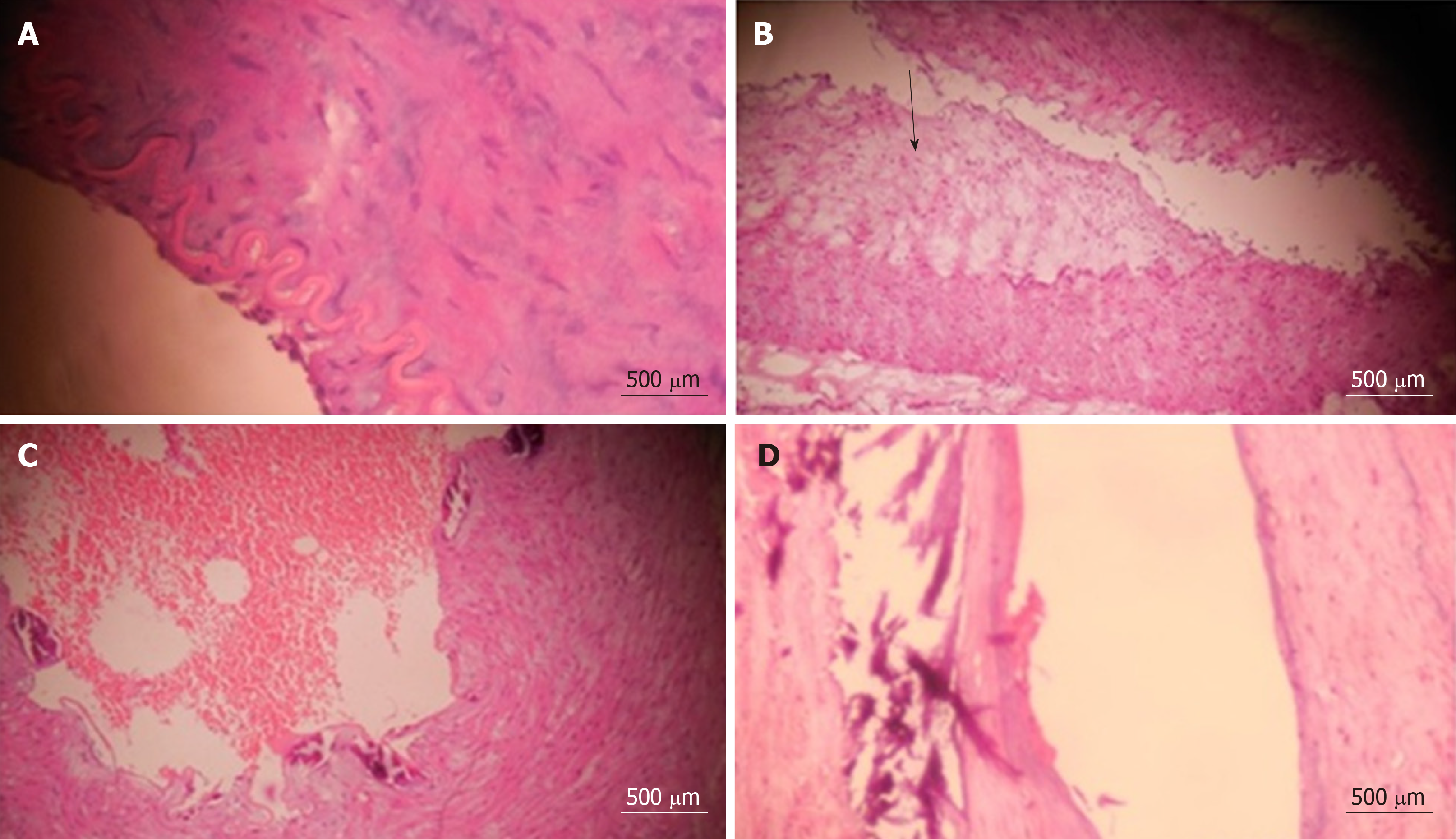
Figure 1 Typical histopathological appearances of all the characteristics of abnormal veins.
A: Histopathology of splenic vein showing medial hypertrophy (Hematoxylin and eosin 40 ×, length of bar 500 µm); B: Histopathology of splenic vein showing intimal fibrosis (arrow) (Hematoxylin and eosin 40 ×, length of bar 500 µm); C: Histopathology of splenic vein showing splenic venous thrombosis (Hematoxylin and eosin 40 ×, length of bar 500 µm); D: Histopathology of splenic vein showing splenic venous wall calcification (Hematoxylin and eosin 40 ×, length of bar 500 µm).
- Citation: Gupta S, Pottakkat B, Verma SK, Kalayarasan R, Chandrasekar A S, Pillai AA. Pathological abnormalities in splenic vasculature in non-cirrhotic portal hypertension: Its relevance in the management of portal hypertension. World J Gastrointest Surg 2020; 12(1): 1-8
- URL: https://www.wjgnet.com/1948-9366/full/v12/i1/1.htm
- DOI: https://dx.doi.org/10.4240/wjgs.v12.i1.1


