Copyright
©The Author(s) 2019.
World J Gastrointest Surg. Sep 27, 2019; 11(9): 373-380
Published online Sep 27, 2019. doi: 10.4240/wjgs.v11.i9.373
Published online Sep 27, 2019. doi: 10.4240/wjgs.v11.i9.373
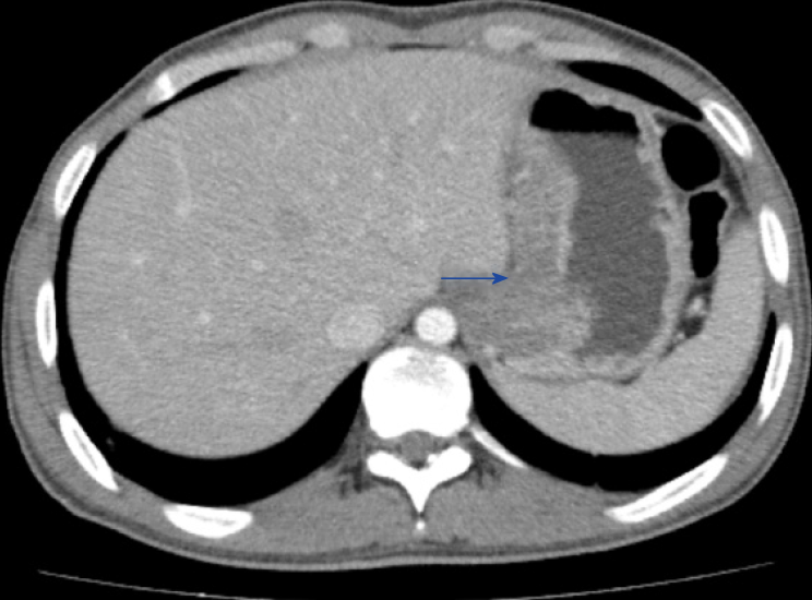
Figure 4 Computed tomography coronal section shows dilated and fluid-filled distal esophagus with significant mural thickening of the distal esophagus involving the gastro-esophageal junction and extending in region of the cardia.
- Citation: Khan MS, Maan MHA, Sohail AH, Memon WA. Primary esophageal tuberculosis mimicking esophageal carcinoma on computed tomography: A case report. World J Gastrointest Surg 2019; 11(9): 373-380
- URL: https://www.wjgnet.com/1948-9366/full/v11/i9/373.htm
- DOI: https://dx.doi.org/10.4240/wjgs.v11.i9.373


