Copyright
©The Author(s) 2019.
World J Gastrointestinal Surgery. Apr 27, 2019; 11(4): 229-236
Published online Apr 27, 2019. doi: 10.4240/wjgs.v11.i4.229
Published online Apr 27, 2019. doi: 10.4240/wjgs.v11.i4.229
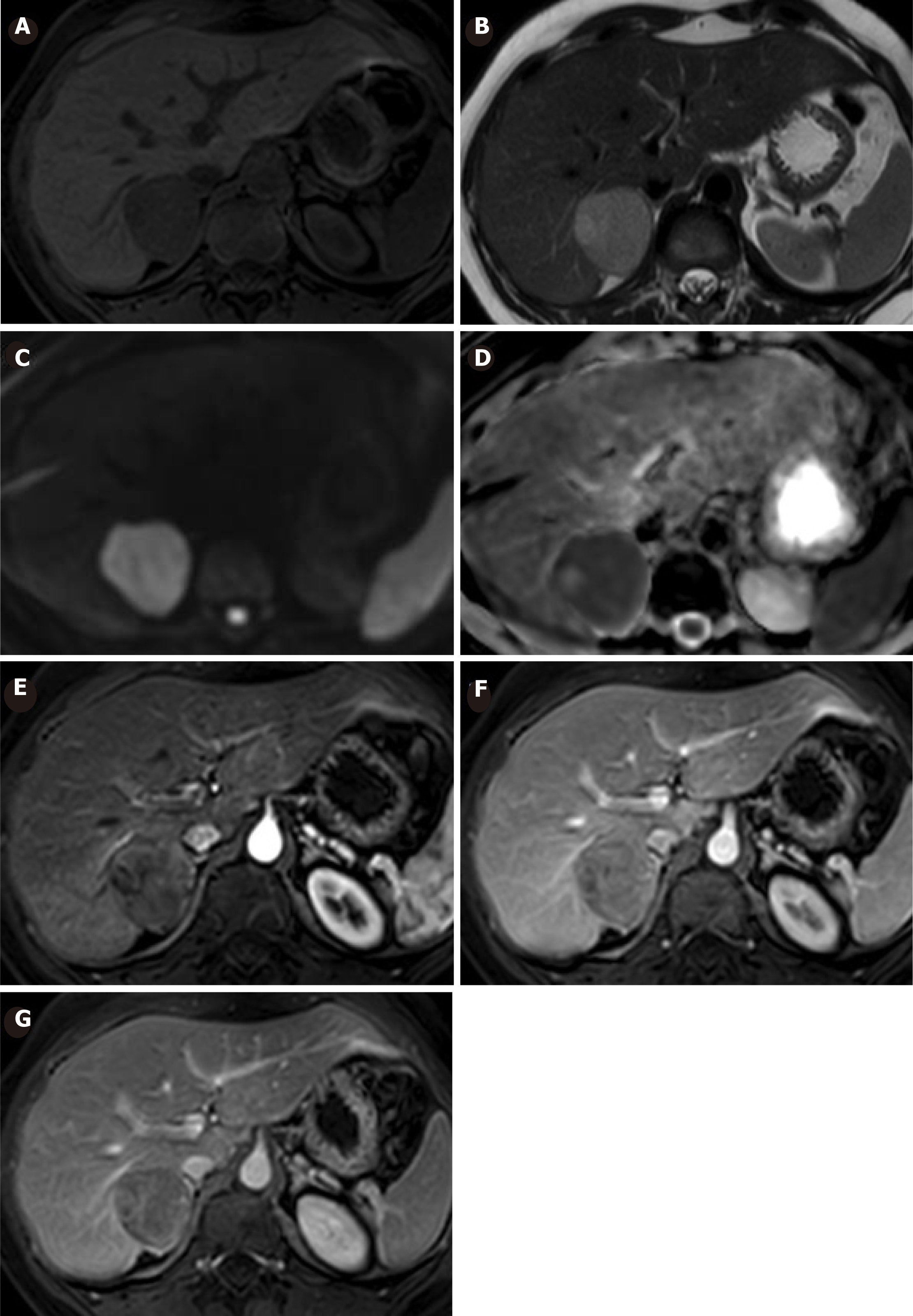
Figure 2 Magnetic resonance imaging is showing a decreased size of the lesion in follow-up.
A: Is hypointense in T2 with an edge of greater signal; B: Is iso-hyperintense in T1; C: It does not show definite evidence of enhancement after administration of the EV contrast in C: Arterial; D: Portal; E: Late phase. These findings could be related to a hepatocellular adenoma with intralesional hemorrhagic changes.
- Citation: Glinka J, Clariá RS, Fratanoni E, Spina J, Mullen E, Ardiles V, Mazza O, Pekolj J, de Santibañes M, de Santibañes E. Malignant transformation of hepatocellular adenoma in a young female patient after ovulation induction fertility treatment: A case report. World J Gastrointestinal Surgery 2019; 11(4): 229-236
- URL: https://www.wjgnet.com/1948-9366/full/v11/i4/229.htm
- DOI: https://dx.doi.org/10.4240/wjgs.v11.i4.229


