Copyright
©The Author(s) 2019.
World J Gastrointest Surg. Feb 27, 2019; 11(2): 85-92
Published online Feb 27, 2019. doi: 10.4240/wjgs.v11.i2.85
Published online Feb 27, 2019. doi: 10.4240/wjgs.v11.i2.85
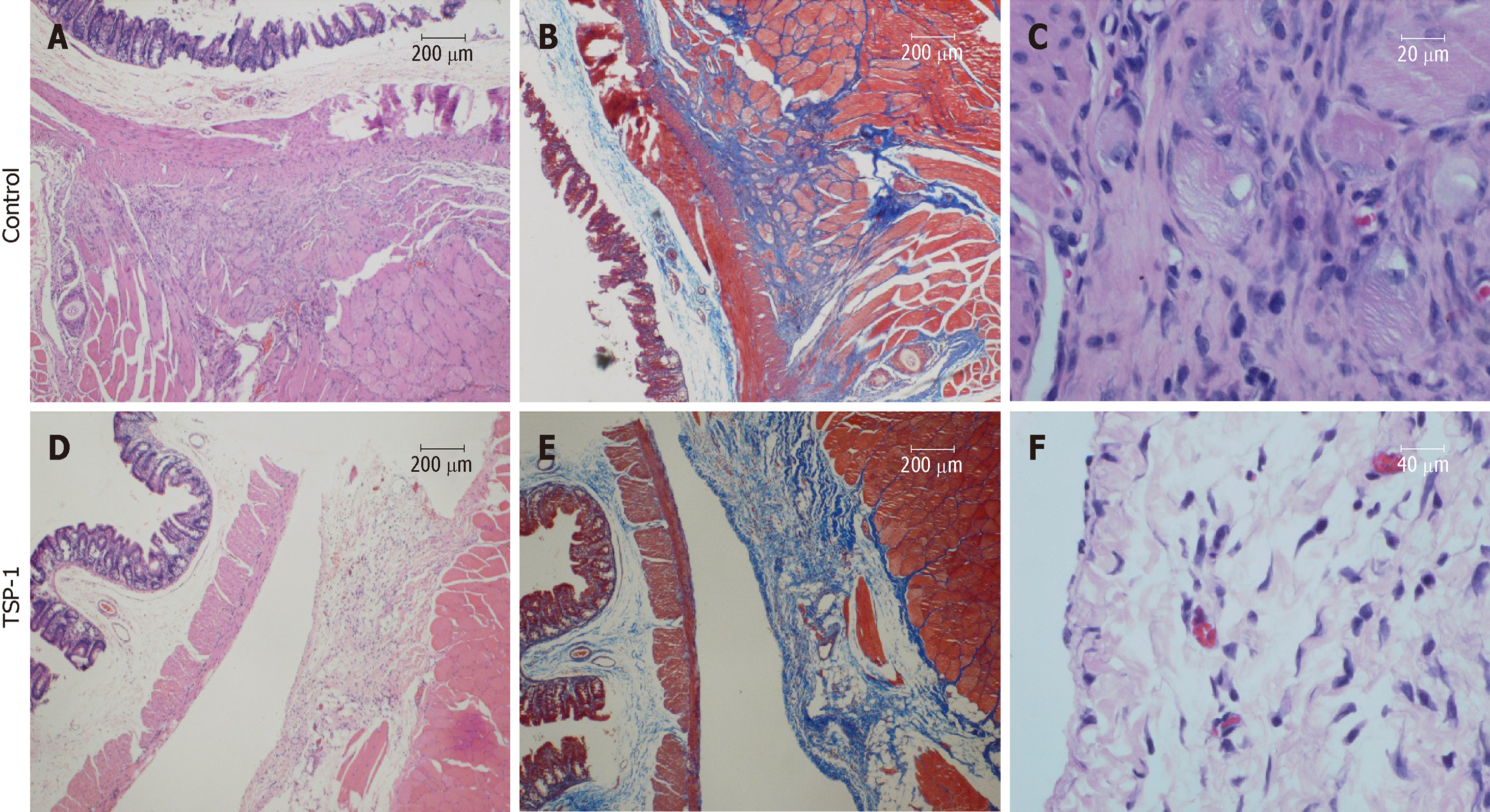
Figure 3 Representative histological sections of cecum with adhesion bands.
A: The muscularis propria is tightly adhered to the skeletal muscle by thick fibrotic tissue and infiltration of mononuclear cells (HE stain, 40 ×); B: The Masson trichrome stain indicates the intersecting of collagen-rich soft-tissue bands formed between the muscle layers (40 ×); C: HE stain shows the proliferative fibroblasts, increased collagen deposition and degenerative skeletal muscles (400 ×); D: Overexpressed TSP-1 on the injured cecum improved the formation of adhesion by separating the intestinal wall and skeletal muscle with a thin layer of loose connective tissue (HE, 40 ×); E: Masson trichrome stain identifies the loose connective tissue beside the skeletal muscle; F: Significantly fewer mononuclear cells infiltration in the loose connective tissue (HE, 400 ×). TSP-1: Thrombospondin 1.
- Citation: Tai YS, Jou IM, Jung YC, Wu CL, Shiau AL, Chen CY. In vivo expression of thrombospondin-1 suppresses the formation of peritoneal adhesion in rats. World J Gastrointest Surg 2019; 11(2): 85-92
- URL: https://www.wjgnet.com/1948-9366/full/v11/i2/85.htm
- DOI: https://dx.doi.org/10.4240/wjgs.v11.i2.85


