Copyright
©The Author(s) 2019.
World J Gastrointest Surg. Feb 27, 2019; 11(2): 62-84
Published online Feb 27, 2019. doi: 10.4240/wjgs.v11.i2.62
Published online Feb 27, 2019. doi: 10.4240/wjgs.v11.i2.62
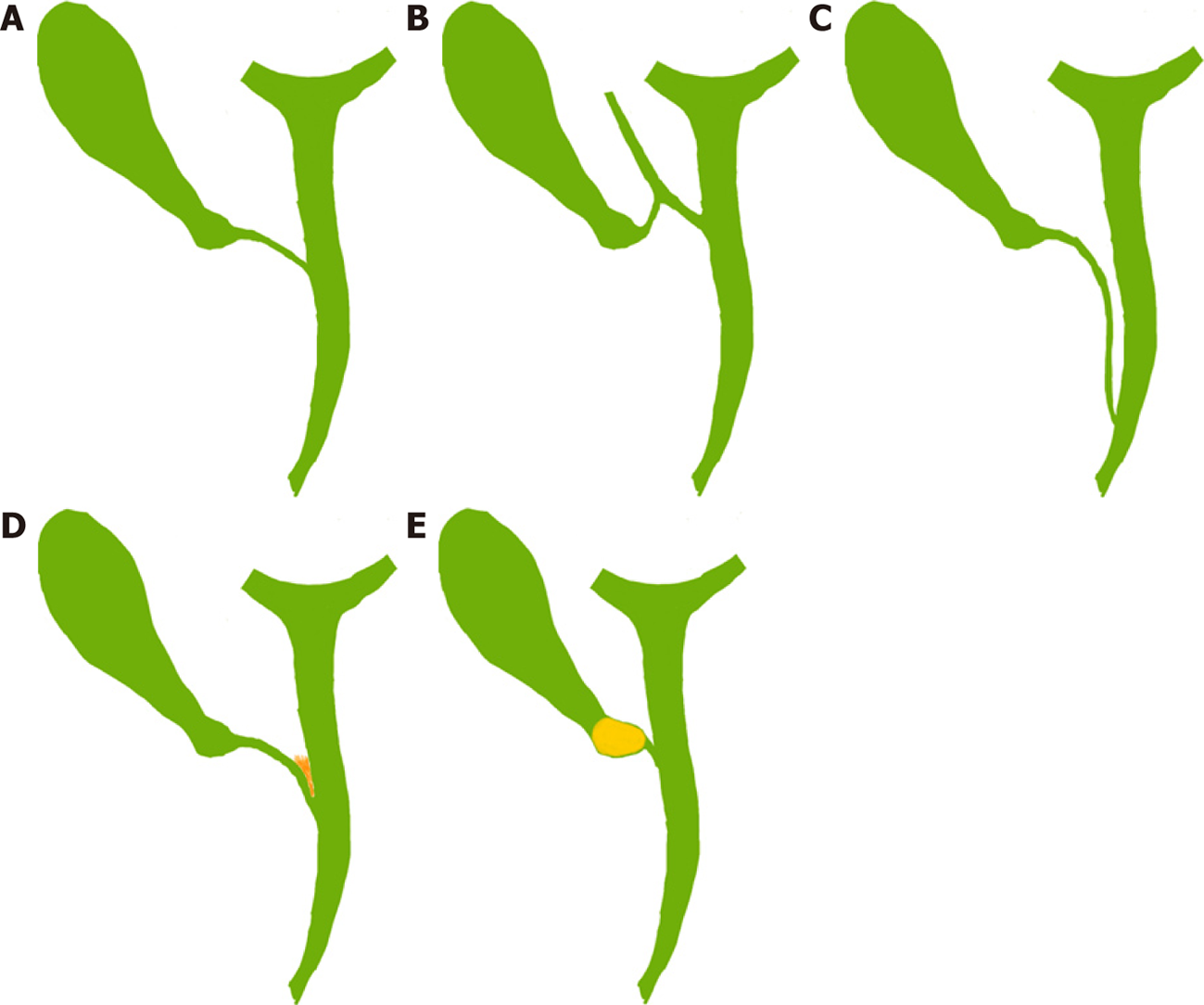
Figure 6 Cystic ductal variations, anatomical and pathological.
A: Normal pattern with angular insertion; B: Cystic duct insertion in aberrant right hepatic (sectional) duct; C: Cystic duct - parallel course. Cystic duct may be quiet long and may join the common hepatic duct (CHD) near ampulla; D: Cystic ductal fusion with the CHD due to inflammation; E: Short/effaced cystic duct due to impacted stone in the gallbladder neck. In both situations (D and E), CHD would be at risk of injury during dissection especially when the surgeon tries to expose the cystic duct-common bile duct junction.
- Citation: Gupta V, Jain G. Safe laparoscopic cholecystectomy: Adoption of universal culture of safety in cholecystectomy. World J Gastrointest Surg 2019; 11(2): 62-84
- URL: https://www.wjgnet.com/1948-9366/full/v11/i2/62.htm
- DOI: https://dx.doi.org/10.4240/wjgs.v11.i2.62


