Copyright
©The Author(s) 2019.
World J Gastrointest Surg. Feb 27, 2019; 11(2): 101-111
Published online Feb 27, 2019. doi: 10.4240/wjgs.v11.i2.101
Published online Feb 27, 2019. doi: 10.4240/wjgs.v11.i2.101
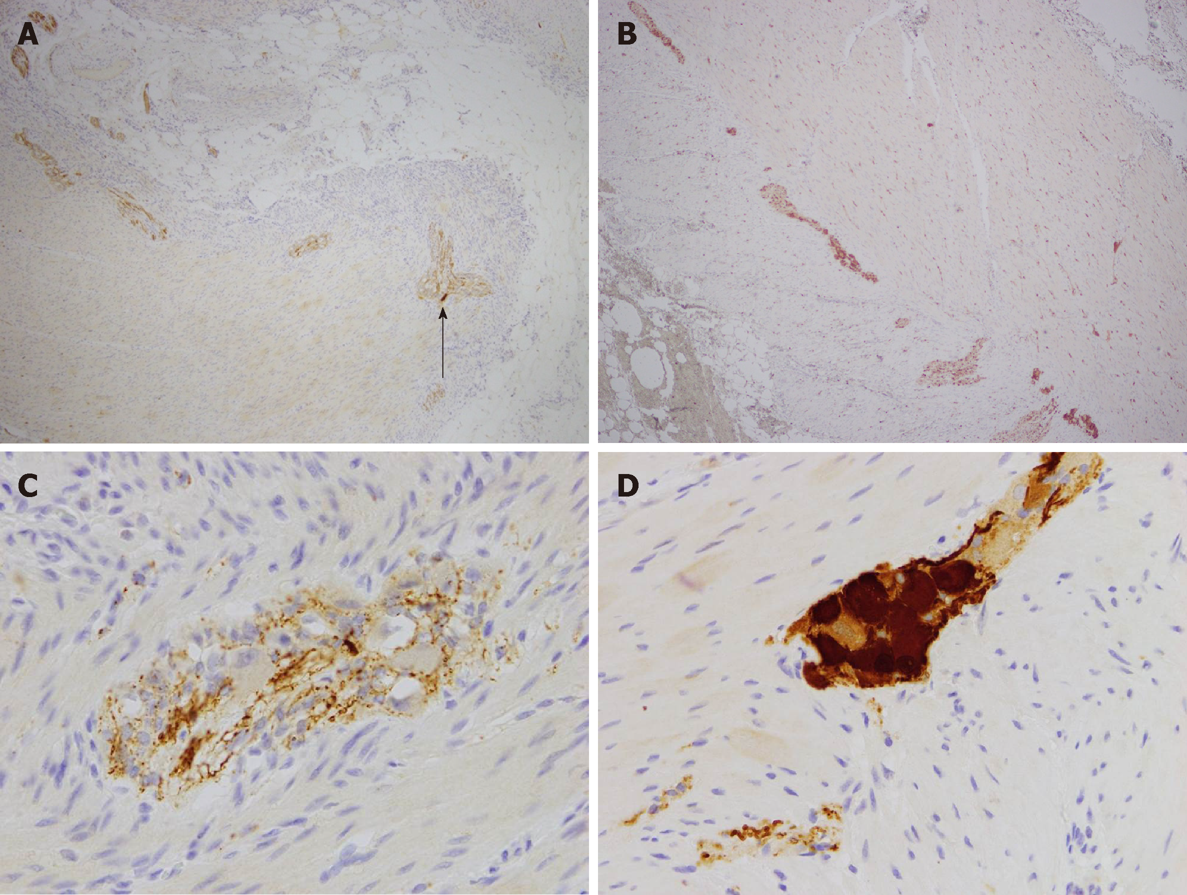
Figure 6 Calretinin staining.
A: Low-power (magnification × 40) calretinin staining of nerve in diseased colon showing one isolated mature ganglion cell (dark brown cell, arrow) and markedly reduced density (0.2/mm2); B: Low-power (magnification × 40) calretinin staining of normal colon showing normal density (5/mm2) of mature ganglion cells (numerous dark brown cells); C: High-power (magnification × 400) calretinin stain in diseased colon showing failure of immature ganglion cells to stain (pale brown cells); D: High-power (magnification × 400) calretinin staining of normal colon showing abundance of mature ganglion cells (dark brown cells).
- Citation: Kwok AM, Still AB, Hart K. Acquired segmental colonic hypoganglionosis in an adult Caucasian male: A case report. World J Gastrointest Surg 2019; 11(2): 101-111
- URL: https://www.wjgnet.com/1948-9366/full/v11/i2/101.htm
- DOI: https://dx.doi.org/10.4240/wjgs.v11.i2.101


