Copyright
©The Author(s) 2019.
World J Gastrointest Surg. Feb 27, 2019; 11(2): 101-111
Published online Feb 27, 2019. doi: 10.4240/wjgs.v11.i2.101
Published online Feb 27, 2019. doi: 10.4240/wjgs.v11.i2.101
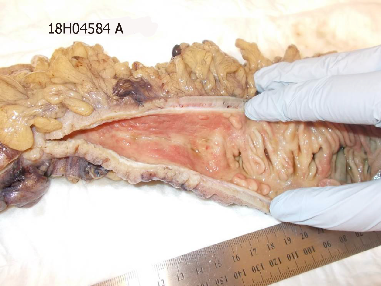
Figure 3 Operative specimen after fixation in formalin.
The segment of colon on the left displays mural thickening, luminal stenosis and exposed submucosa secondary to the mucosa being denuded. There is a sharp transition to relatively normal colon on the right, with intact mucosal folds being observed.
- Citation: Kwok AM, Still AB, Hart K. Acquired segmental colonic hypoganglionosis in an adult Caucasian male: A case report. World J Gastrointest Surg 2019; 11(2): 101-111
- URL: https://www.wjgnet.com/1948-9366/full/v11/i2/101.htm
- DOI: https://dx.doi.org/10.4240/wjgs.v11.i2.101


