Copyright
©The Author(s) 2019.
World J Gastrointest Surg. Dec 27, 2019; 11(12): 443-448
Published online Dec 27, 2019. doi: 10.4240/wjgs.v11.i12.443
Published online Dec 27, 2019. doi: 10.4240/wjgs.v11.i12.443
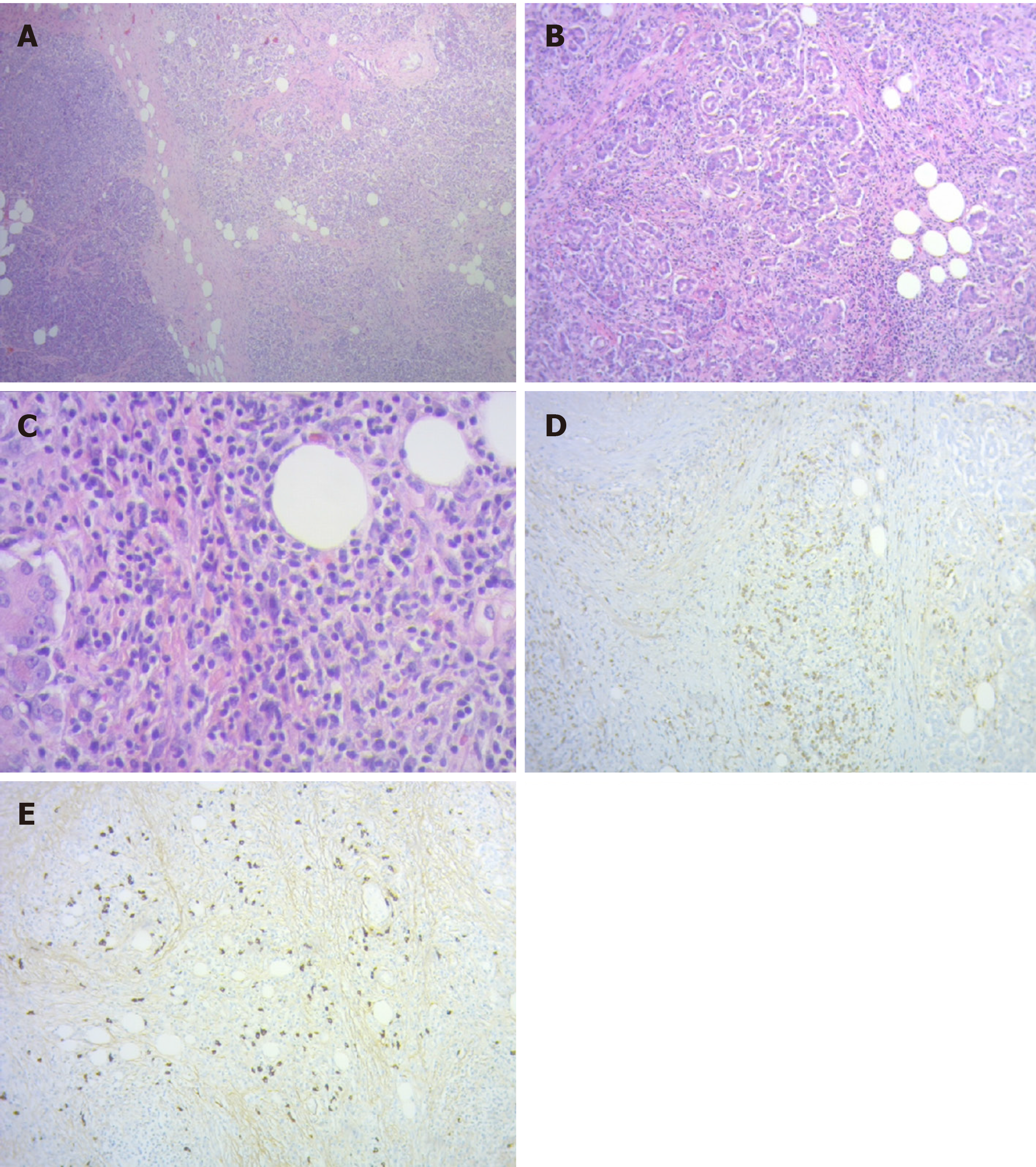
Figure 3 Histopathological analysis of the surgical specimen.
A: Limits between normal pancreatic tissue and carcinoma (hematoxylin and eosin 40 ×); B: Lymphoplasmacytic peritumoral infiltration (hematoxylin and eosin 100 ×); C: Plasmacytic peritumoral infiltration (hematoxylin and eosin 400 ×); D: Immunohistochemistry with IgG positive plasma cells; E: Immunohistochemistry with frequent IgG4 positive plasma cells.
- Citation: Glinka J, Calderón F, de Santibañes M, Hyon SH, Gadano A, Mullen E, Pol M, Spina J, de Santibañes E. Early pancreatic cancer in IgG4-related pancreatic mass: A case report. World J Gastrointest Surg 2019; 11(12): 443-448
- URL: https://www.wjgnet.com/1948-9366/full/v11/i12/443.htm
- DOI: https://dx.doi.org/10.4240/wjgs.v11.i12.443


