Copyright
©The Author(s) 2019.
World J Gastrointest Surg. Dec 27, 2019; 11(12): 443-448
Published online Dec 27, 2019. doi: 10.4240/wjgs.v11.i12.443
Published online Dec 27, 2019. doi: 10.4240/wjgs.v11.i12.443
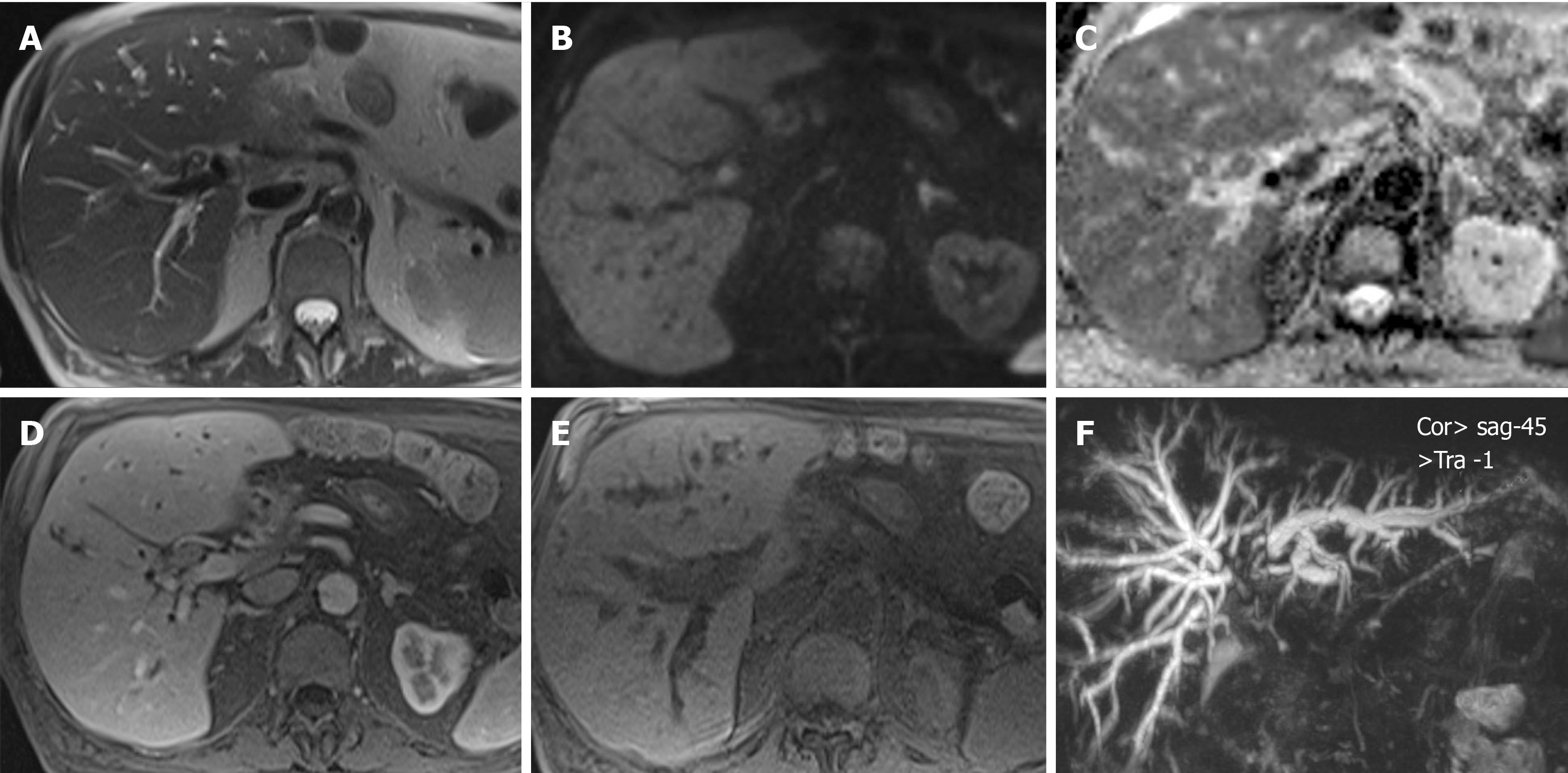
Figure 2 Magnetic resonance cholangio-pancreatography showing parietal concentric ingrowth of the main bile duct with stenosis from the hepatic carrefour up to the hepaticojejunostomy with dilatation of intrahepatic bile ducts.
A: Axial T2 with concentric parietal growth; B: Diffusion-weighted imaging b800. Hyperintensity; C: Apparent diffusion coefficient with hyperintensity; D: Axial T1 without contrast; E: Axial in a portal phase; F: Three dimensional cholangiography showing dilation of intrahepatic ducts superior to the hepatic carrefour with stenosis from the carrefour up to the anastomosis.
- Citation: Glinka J, Calderón F, de Santibañes M, Hyon SH, Gadano A, Mullen E, Pol M, Spina J, de Santibañes E. Early pancreatic cancer in IgG4-related pancreatic mass: A case report. World J Gastrointest Surg 2019; 11(12): 443-448
- URL: https://www.wjgnet.com/1948-9366/full/v11/i12/443.htm
- DOI: https://dx.doi.org/10.4240/wjgs.v11.i12.443


