Copyright
©The Author(s) 2018.
World J Gastrointest Surg. Nov 27, 2018; 10(8): 90-94
Published online Nov 27, 2018. doi: 10.4240/wjgs.v10.i8.90
Published online Nov 27, 2018. doi: 10.4240/wjgs.v10.i8.90
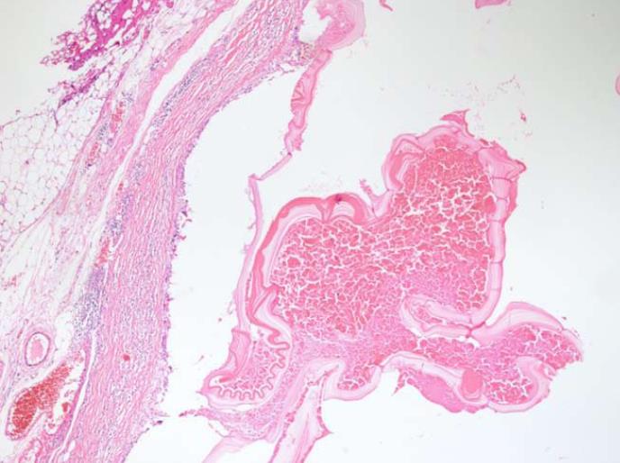
Figure 6 Microscopic appearance of the hydatid cyst tissue stained with hematoxylin and eosin.
An acellular membrane of a hydatid cyst is shown here (HE × 40).
- Citation: Akbulut S, Yilmaz M, Alan S, Kolu M, Karadag N. Coexistence of duodenum derived aggressive fibromatosis and paraduodenal hydatid cyst: A case report and review of literature. World J Gastrointest Surg 2018; 10(8): 90-94
- URL: https://www.wjgnet.com/1948-9366/full/v10/i8/90.htm
- DOI: https://dx.doi.org/10.4240/wjgs.v10.i8.90


