Copyright
©The Author(s) 2018.
World Journal of Gastrointestinal Surgery. Aug 27, 2018; 10(5): 49-56
Published online Aug 27, 2018. doi: 10.4240/wjgs.v10.i5.49
Published online Aug 27, 2018. doi: 10.4240/wjgs.v10.i5.49
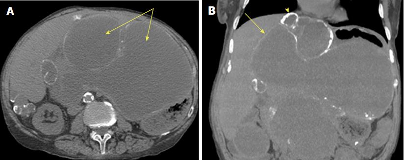
Figure 3 Computed tomography of pseudomyxoma peritonei[26].
This is an axial computed tomography scan. A: Cystic accumulations of mucus (arrows) surrounded by calcified rims; B: A coronal reconstruction representing cystic accumulations in the upper abdomen and the liver.
- Citation: Rizvi SA, Syed W, Shergill R. Approach to pseudomyxoma peritonei. World Journal of Gastrointestinal Surgery 2018; 10(5): 49-56
- URL: https://www.wjgnet.com/1948-9366/full/v10/i5/49.htm
- DOI: https://dx.doi.org/10.4240/wjgs.v10.i5.49


