Copyright
©The Author(s) 2018.
World J Gastrointest Surg. Jan 27, 2018; 10(1): 1-5
Published online Jan 27, 2018. doi: 10.4240/wjgs.v10.i1.1
Published online Jan 27, 2018. doi: 10.4240/wjgs.v10.i1.1
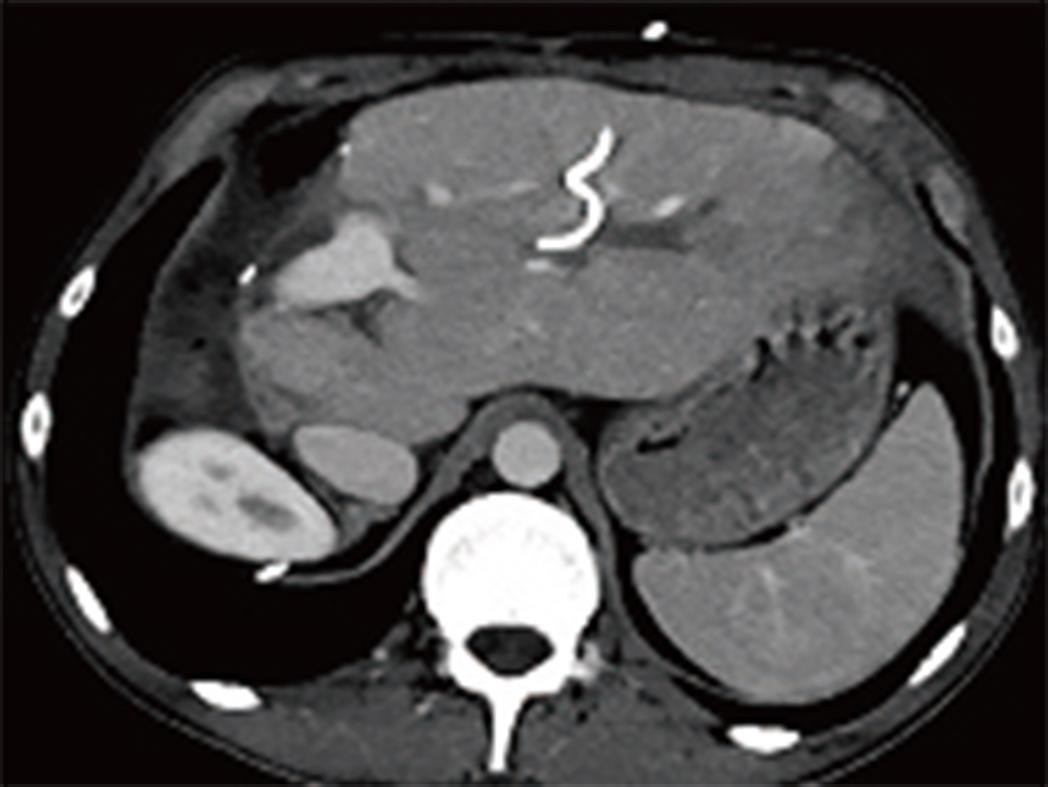
Figure 4 Postoperative axial phase multidetector computerized tomography image.
Extended right hepatectomy involving the right lobe and segment IV is performed remaining the segment II and III. Segment III bile duct is dilated (white arrow) and an external biliary drainage catheter is seen inside the bile duct (black arrow).
- Citation: Akbulut S, Cicek E, Kolu M, Sahin TT, Yilmaz S. Associating liver partition and portal vein ligation for staged hepatectomy for extensive alveolar echinococcosis: First case report in the literature. World J Gastrointest Surg 2018; 10(1): 1-5
- URL: https://www.wjgnet.com/1948-9366/full/v10/i1/1.htm
- DOI: https://dx.doi.org/10.4240/wjgs.v10.i1.1


