Copyright
©The Author(s) 2018.
World J Gastrointest Surg. Jan 27, 2018; 10(1): 1-5
Published online Jan 27, 2018. doi: 10.4240/wjgs.v10.i1.1
Published online Jan 27, 2018. doi: 10.4240/wjgs.v10.i1.1
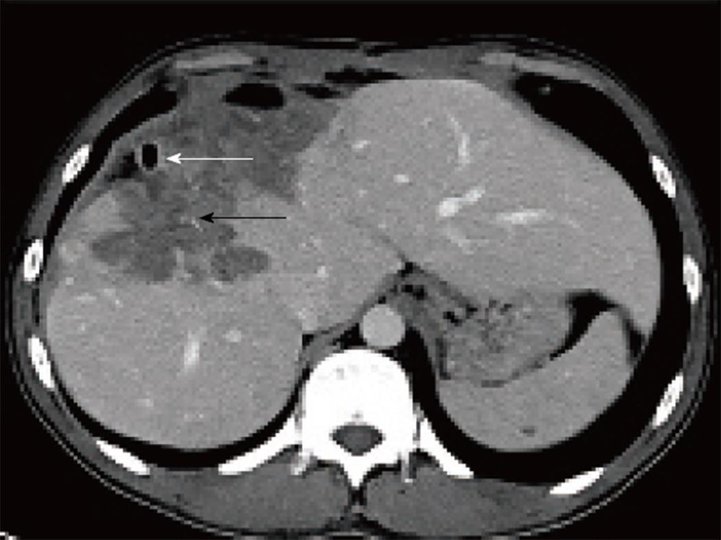
Figure 2 Preoperative axial venous phase multidetector computerized tomography image.
A percutaneous drainage catheter to drain the infected content of the lesion is seen (white arrow). Furthermore, it observed that the cystic lesion causes a retraction in the hepatic capsule and shows invasion to surrounding tissue with millimetric amorphous calcification foci (black arrow).
- Citation: Akbulut S, Cicek E, Kolu M, Sahin TT, Yilmaz S. Associating liver partition and portal vein ligation for staged hepatectomy for extensive alveolar echinococcosis: First case report in the literature. World J Gastrointest Surg 2018; 10(1): 1-5
- URL: https://www.wjgnet.com/1948-9366/full/v10/i1/1.htm
- DOI: https://dx.doi.org/10.4240/wjgs.v10.i1.1


