Copyright
©The Author(s) 2015.
World J Gastrointest Surg. May 27, 2015; 7(5): 78-81
Published online May 27, 2015. doi: 10.4240/wjgs.v7.i5.78
Published online May 27, 2015. doi: 10.4240/wjgs.v7.i5.78
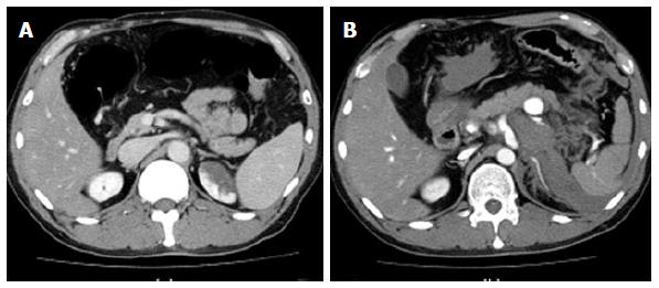
Figure 1 Enhanced computed tomography demonstrated no splenic artery aneurysm two weeks prior to the hospital visit (A) and a 20 mm aneurysm of the splenic artery and hematoma accumulated around the retroperitoneum (B).
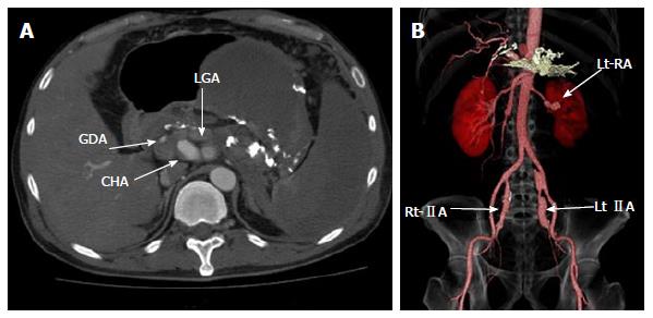
Figure 2 Enhanced computed tomography demonstrated six splanchnic artery aneurysms.
GDA: Gastric duodenal artery; LGA: Left gastric artery; CHA: Common hepatic artery; Lt-RA: Left renal artery; Rt-IIA: Right internal iliac artery; Lt-IIA: Left internal iliac artery.
Figure 3 Celiac angiography revealed aneurysms in the splenic, left gastric, and common hepatic arteries (A), splenic angiography demonstrated extravasation from the splenic artery aneurysm (arrow) (B), and successful microcoil embolization of the ruptured splenic artery aneurysm with complete cessation of flow within the aneurysm (arrow) (C).
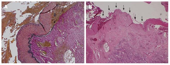
Figure 4 Histopathology revealed an adventitial-medial junction created by separation of the media from the adventitia and organized thrombi deposited at the site.
A: Organized thrombi were deposited at the adventitial-medial junction created by the incipient separation of the media from the adventitia (arrow). Elastica Van Gieson stain; magnification × 4; B: Mediolysis can involve the entire medial muscle with preservation of the intima and internal elastica (arrow). The overlying adventitial-medial junction is suffused with fibrin. Hematoxylin Eosin stain; Magnification × 2.
- Citation: Matsuda Y, Sakamoto K, Nishino E, Kataoka N, Yamaguchi T, Tomita M, Kazi A, Shinozaki M, Makimoto S. Pancreatectomy and splenectomy for a splenic aneurysm associated with segmental arterial mediolysis. World J Gastrointest Surg 2015; 7(5): 78-81
- URL: https://www.wjgnet.com/1948-9366/full/v7/i5/78.htm
- DOI: https://dx.doi.org/10.4240/wjgs.v7.i5.78


