Copyright
©The Author(s) 2023.
World J Gastrointest Surg. Dec 27, 2023; 15(12): 2954-2961
Published online Dec 27, 2023. doi: 10.4240/wjgs.v15.i12.2954
Published online Dec 27, 2023. doi: 10.4240/wjgs.v15.i12.2954
Figure 1 Multiparameter monitor.
Monitor was turned on with parameter detection: electrocardiogram, noninvasive blood pressure and pulse oximetry.
Figure 2 Mechanical ventilation monitor.
Monitor was turned off because the patient was breathing spontaneously.
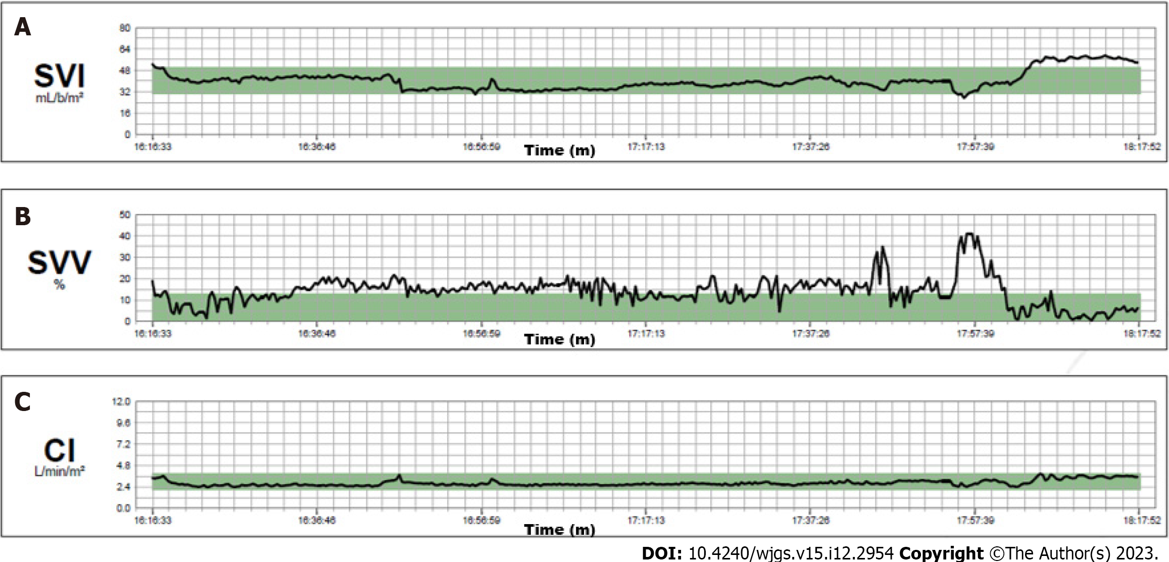
Figure 3 Intraoperative hemodynamic monitoring.
A: Stroke volume index curve; B: Stroke volume variation curve; C: Cardiac index curve. SVI: Stroke volume index curve; SVV: Stroke volume variation curve; CI: Cardiac Index curve.
Figure 4 Intraoperative mean arterial pressure: mean arterial pressure.
MAP curve. Arrows indicate four boluses of ephedrine. MAP: Mean arterial pressure.
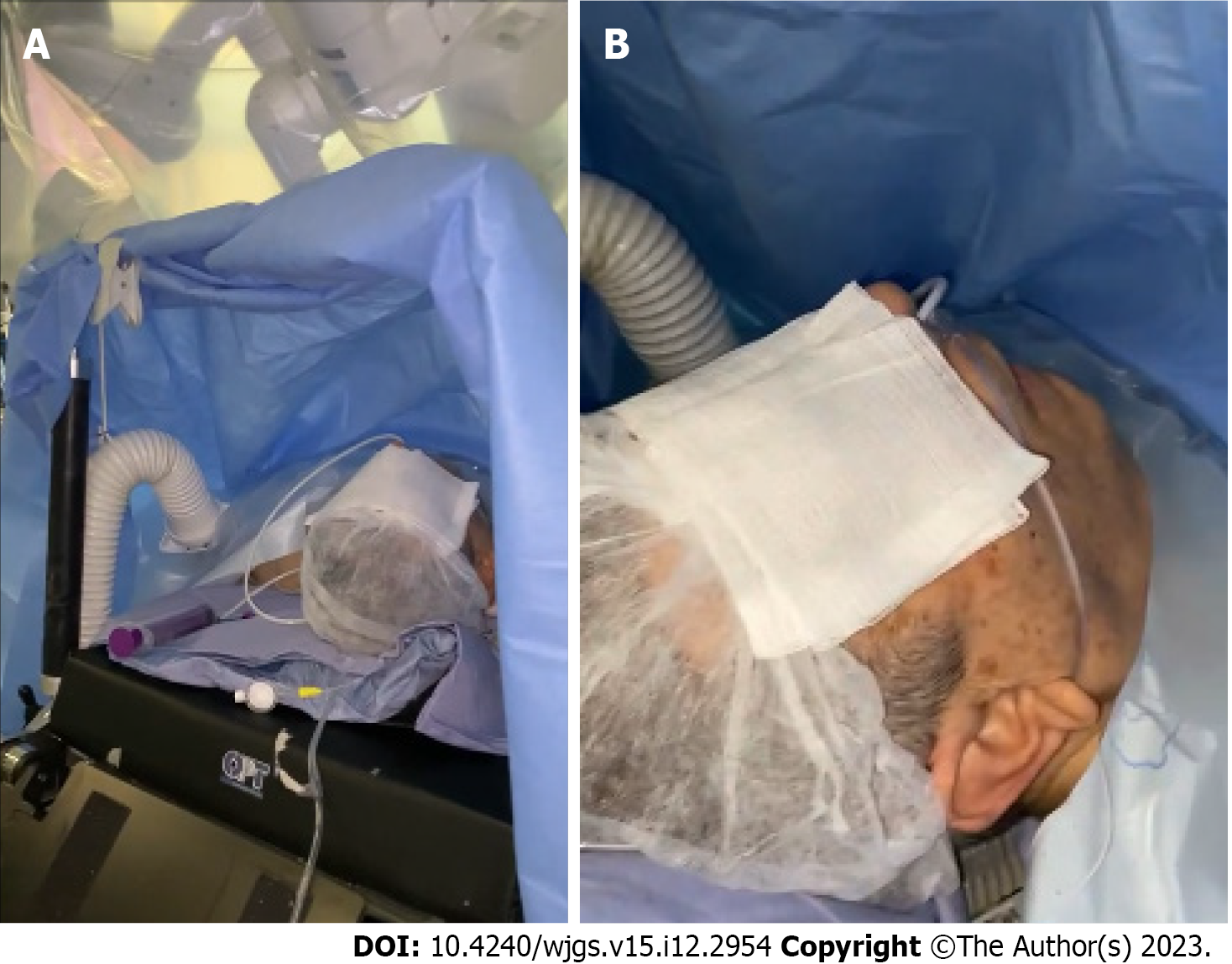
Figure 5 Patient awake in a supine position.
A: The patient was awake and placed in the supine position with 30° reverse Trendelenburg under the robotic arms; B: The patient was awake in the absence of an endotracheal tube.
Figure 6 Trocar positions.
Patient was placed in a supine position with 30° reverse Trendelenburg position. A pneumoperitoneum was created using an open technique through a supraumbilical incision. The other three robotic trocar, right pararectal, left pararectal and left flank, and two laparoscopic 12-mm trocar assistants, right and left iliac fossa, were inserted under direct vision.
- Citation: Delvecchio A, Pavone G, Conticchio M, Piacente C, Varvara M, Ferraro V, Stasi M, Casella A, Filippo R, Tedeschi M, Pullano C, Inchingolo R, Delmonte V, Memeo R. Awake robotic liver surgery: A case report. World J Gastrointest Surg 2023; 15(12): 2954-2961
- URL: https://www.wjgnet.com/1948-9366/full/v15/i12/2954.htm
- DOI: https://dx.doi.org/10.4240/wjgs.v15.i12.2954


